Uppladdningar av Jadwiga Krusnell
Hoppa till navigering
Hoppa till sök
Den här specialsidan visar alla filer som laddats upp.
| Datum | Namn | Miniatyrbild | Storlek (byte) | Beskrivning | Versioner |
|---|---|---|---|---|---|
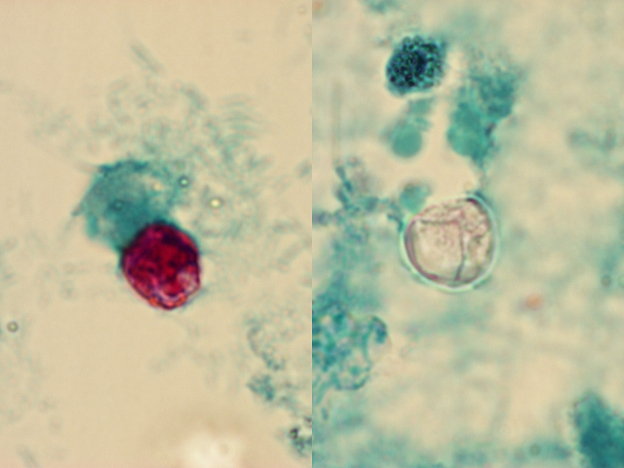
| 14 mars 2012 kl. 15.47 | Cyclospora Bild 2. 100x.jpg (fil) |  |
233 kbyte | 1 | |
| 14 mars 2012 kl. 15.43 | Cyclospora crop Bild 1.jpg (fil) |  |
69 kbyte | 1 | |
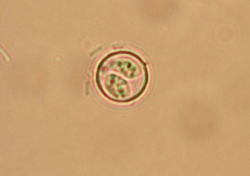
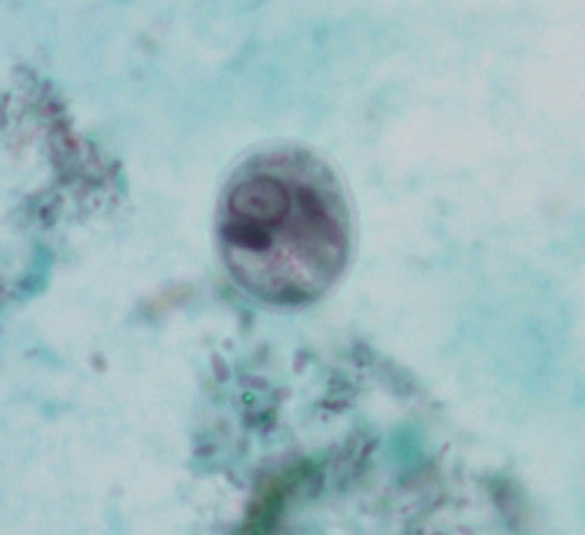
| 6 mars 2012 kl. 16.34 | Cyclospora Bild 4 sporulerad oocysta crop.jpg (fil) |  |
36 kbyte | 1 | |
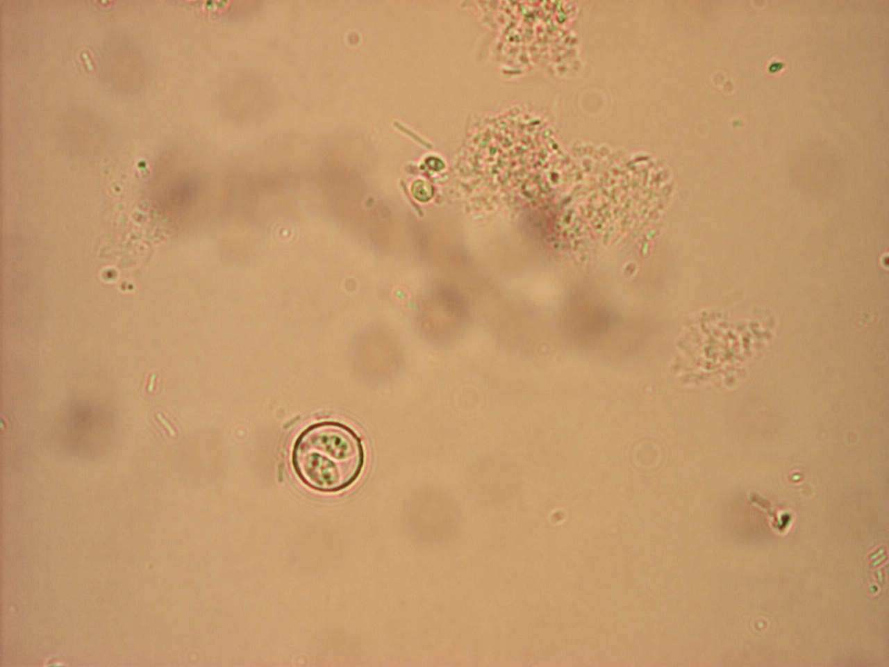
| 6 mars 2012 kl. 16.29 | Cyclospora Bild 4 sporulerad oocysta.jpeg (fil) |  |
226 kbyte | 1 | |
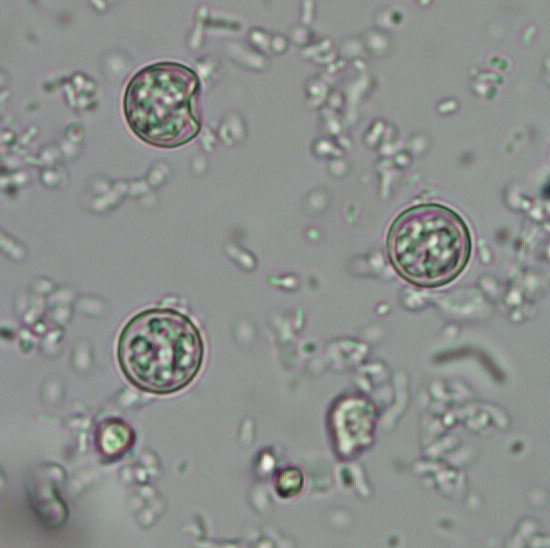
| 6 mars 2012 kl. 16.27 | Cyclospora bild 3.jpeg (fil) |  |
157 kbyte | 1 | |
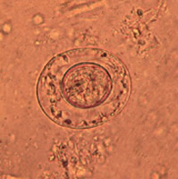
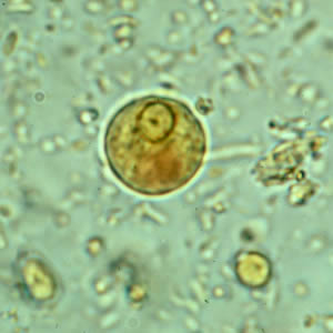
| 6 mars 2012 kl. 15.32 | Isospora bild 2.jpg (fil) |  |
675 kbyte | OOcysta från Isospora (Cycloisospora) hominis. Foto: Marianne Lebbad SMI | 1 |
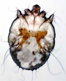
| 22 september 2011 kl. 14.32 | Skabb. Foto.jpg (fil) |  |
42 kbyte | Foto: Lill-Marie Persson, Kärnsjukhuset i Skövde | 1 |
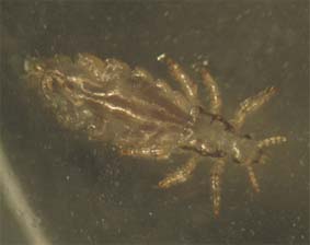
| 22 september 2011 kl. 14.30 | Lus Foto CT.jpg (fil) |  |
34 kbyte | CT SMI | 1 |
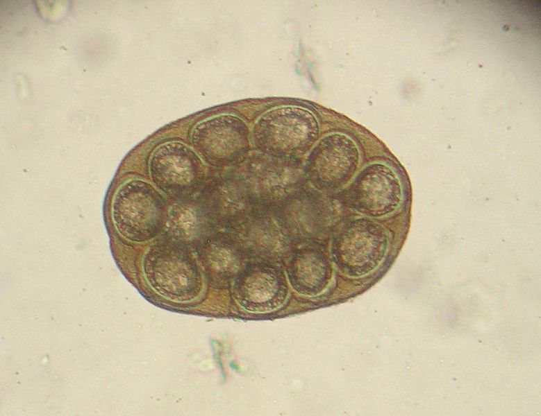
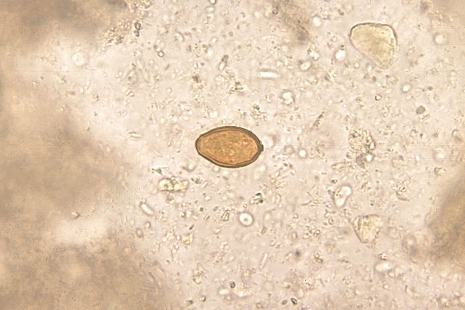
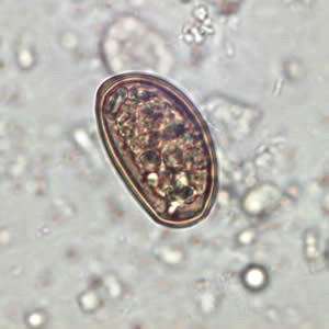
| 20 september 2011 kl. 14.08 | H.diminutaÄggML-a.jpg (fil) |  |
283 kbyte | Foto:marianne Lebbad, Smittskyddsinstitutet | 1 |
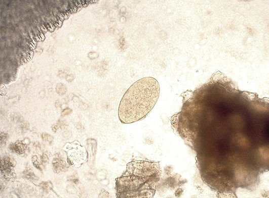
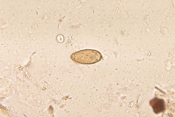
| 15 september 2011 kl. 15.16 | H nana eggB. G.dept.publ.health.jpg (fil) |  |
18 kbyte | Georgia Dept. Publ. Health via Wikimedia Commons | 1 |
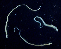
| 15 september 2011 kl. 15.14 | H nana adultF. cdc.jpg (fil) |  |
15 kbyte | CDC via Wikimedia Commons | 1 |
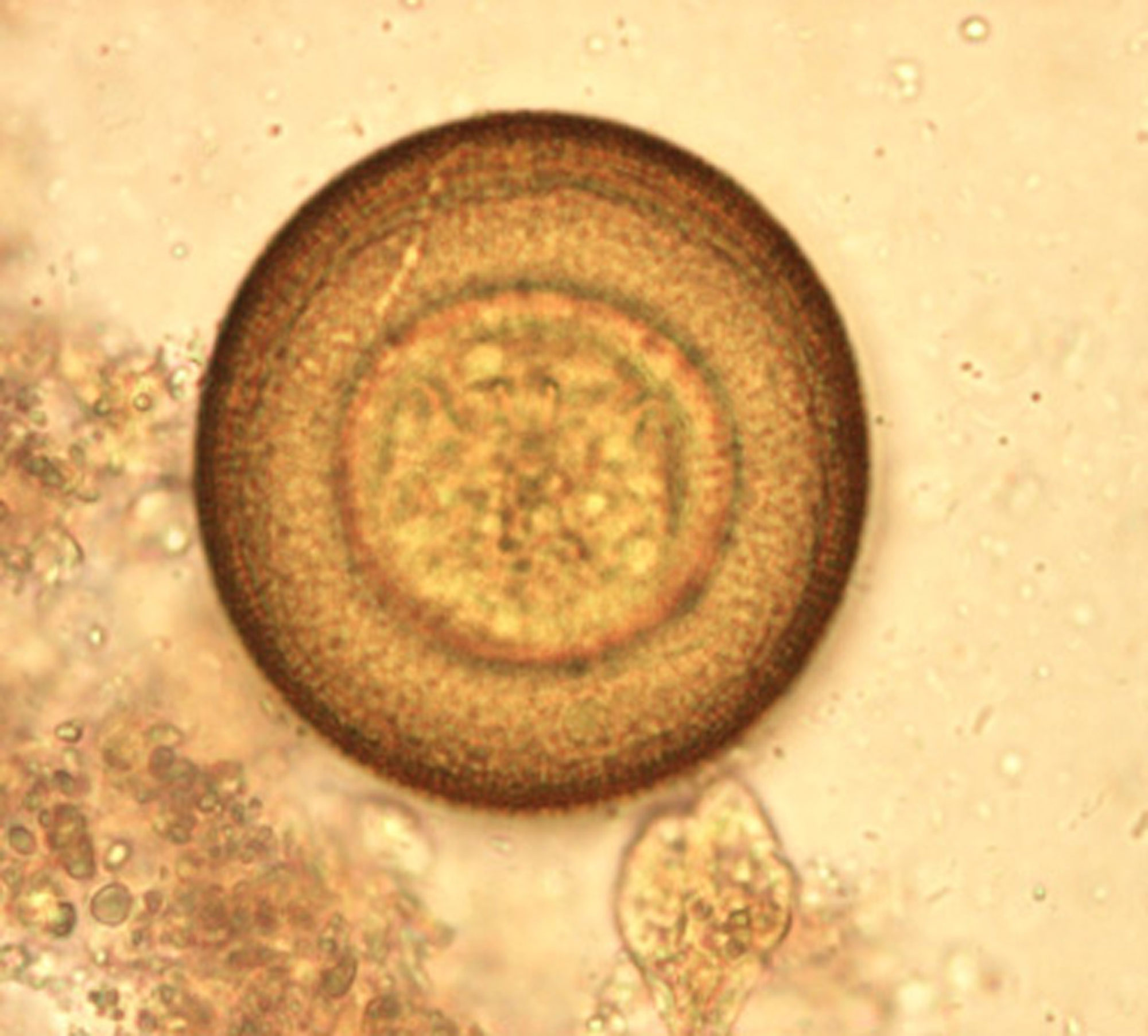
| 14 september 2011 kl. 14.19 | 779px-Dipylidium caninum ovum 1.jpg (fil) |  |
53 kbyte | Foto Joel Mills via Wikimedia Commons | 2 |
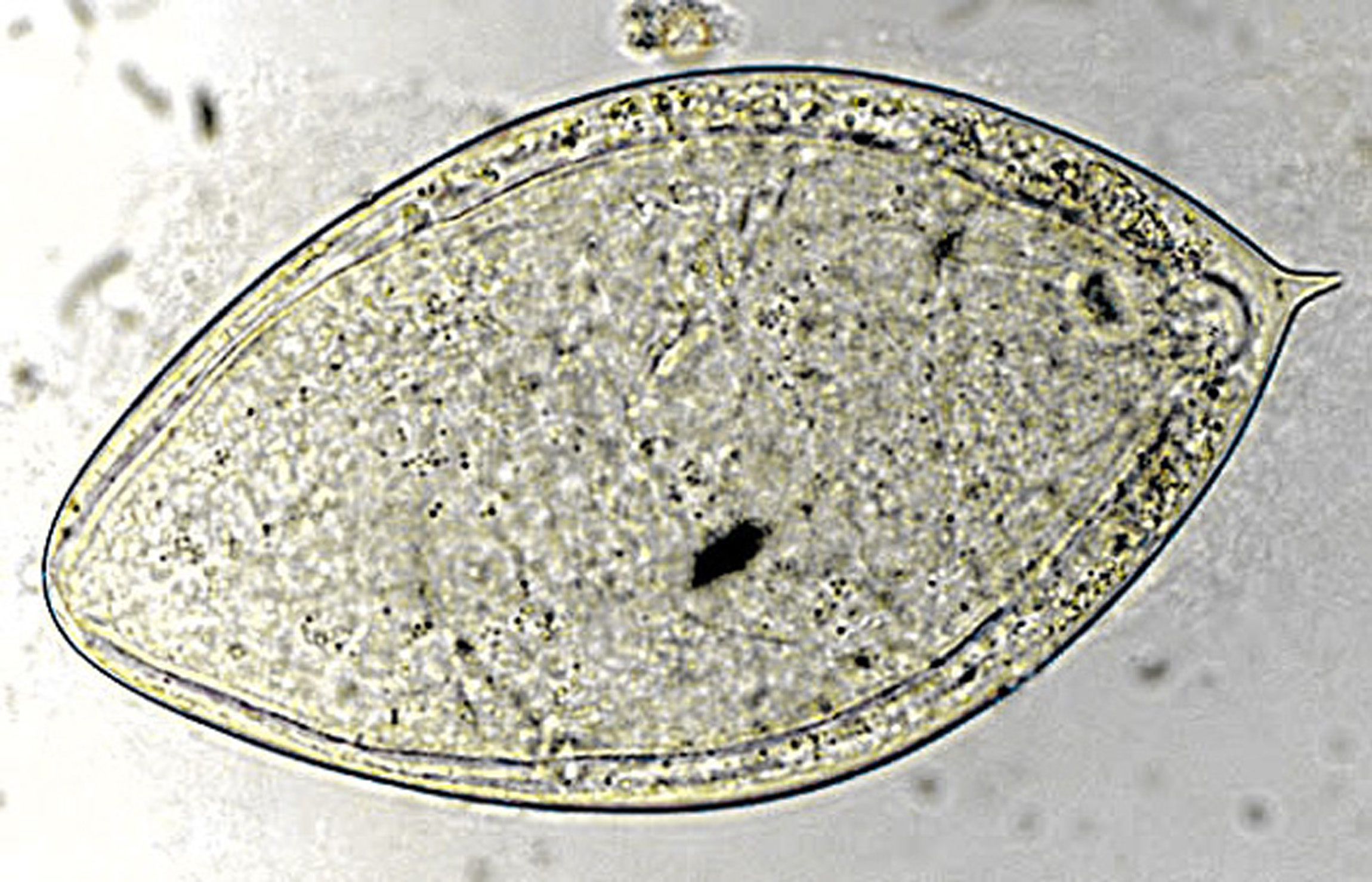
| 8 september 2011 kl. 16.12 | Heterophyes egg.jpg (fil) |  |
64 kbyte | Foto: CDC via Wikimedia Commons | 1 |
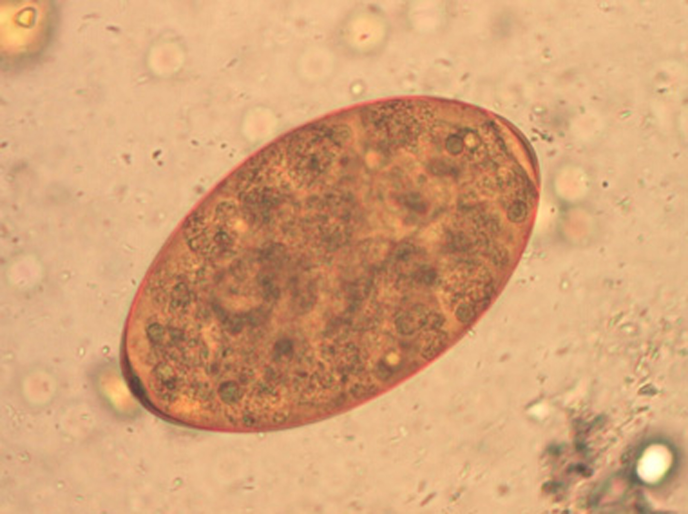
| 8 september 2011 kl. 15.16 | Egg of Fasciola hepatica 08G0041 lores.jpg (fil) |  |
48 kbyte | Foto: CDC via Wikimedia Commons | 1 |
| 8 september 2011 kl. 15.05 | FasciolaÄggML.jpg (fil) |  |
417 kbyte | Foto: Marianne Lebbad, Smittskyddsinstitutet | 1 |
| 8 september 2011 kl. 11.16 | S.mansoniÄggAM.jpg (fil) |  |
547 kbyte | Foto: Anders Magnusson, Smittskyddsinstitutet | 1 |
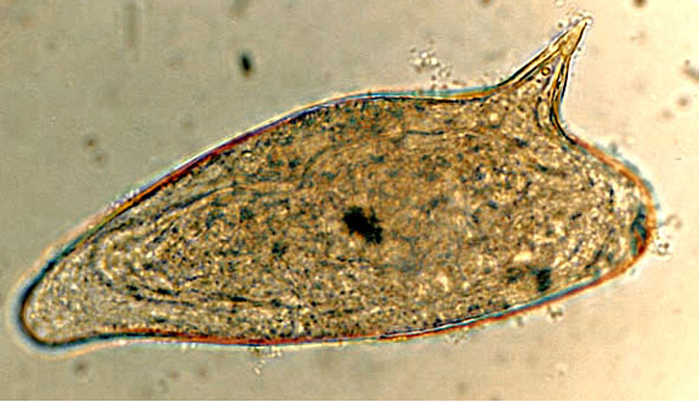
| 8 september 2011 kl. 11.12 | S.HaemobiumÄggAM.jpg (fil) |  |
673 kbyte | Foto: Anders Magnusson, Smittskyddsinstitutet | 1 |
| 7 september 2011 kl. 12.20 | Clonorchis sinensis egg 06G0049 jpg lores.jpg (fil) |  |
51 kbyte | Foto: CDC via Wikimedia Commons | 1 |
| 7 september 2011 kl. 10.19 | ClonorchOpisthÄggBS-a.jpg (fil) |  |
344 kbyte | Foto: Silvia Botero Kleiven, Smittskyddsinstitutet | 1 |
| 6 september 2011 kl. 15.50 | D dendriticum egg wtmt JCG C.jpg (fil) |  |
29 kbyte | Foto: CDC vie Wikimedia Commons | 1 |
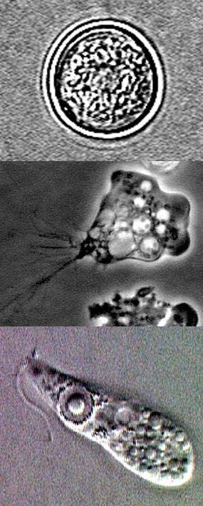
| 5 september 2011 kl. 14.59 | Naegleria fowleri lifecycle stages rotated.jpg (fil) |  |
77 kbyte | Foto: Wiki media commons | 1 |
| 5 september 2011 kl. 14.47 | Naegleria trophA.jpg (fil) |  |
7 kbyte | Foto: Wiki media commons | 1 |
| 5 september 2011 kl. 14.37 | Ehistdisp cyst wtmt.jpg (fil) |  |
14 kbyte | Foto: Wiki media commons | 1 |
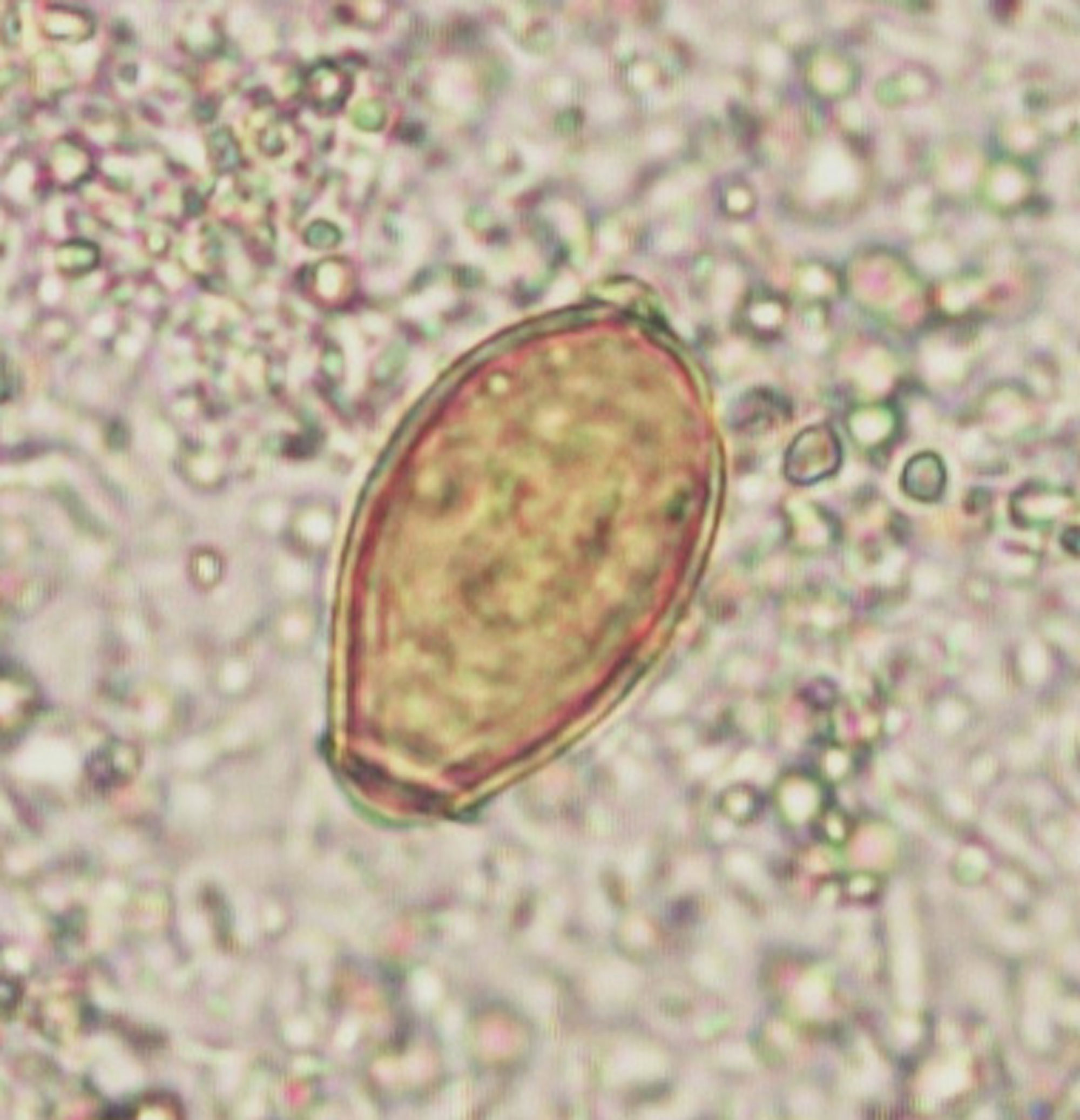
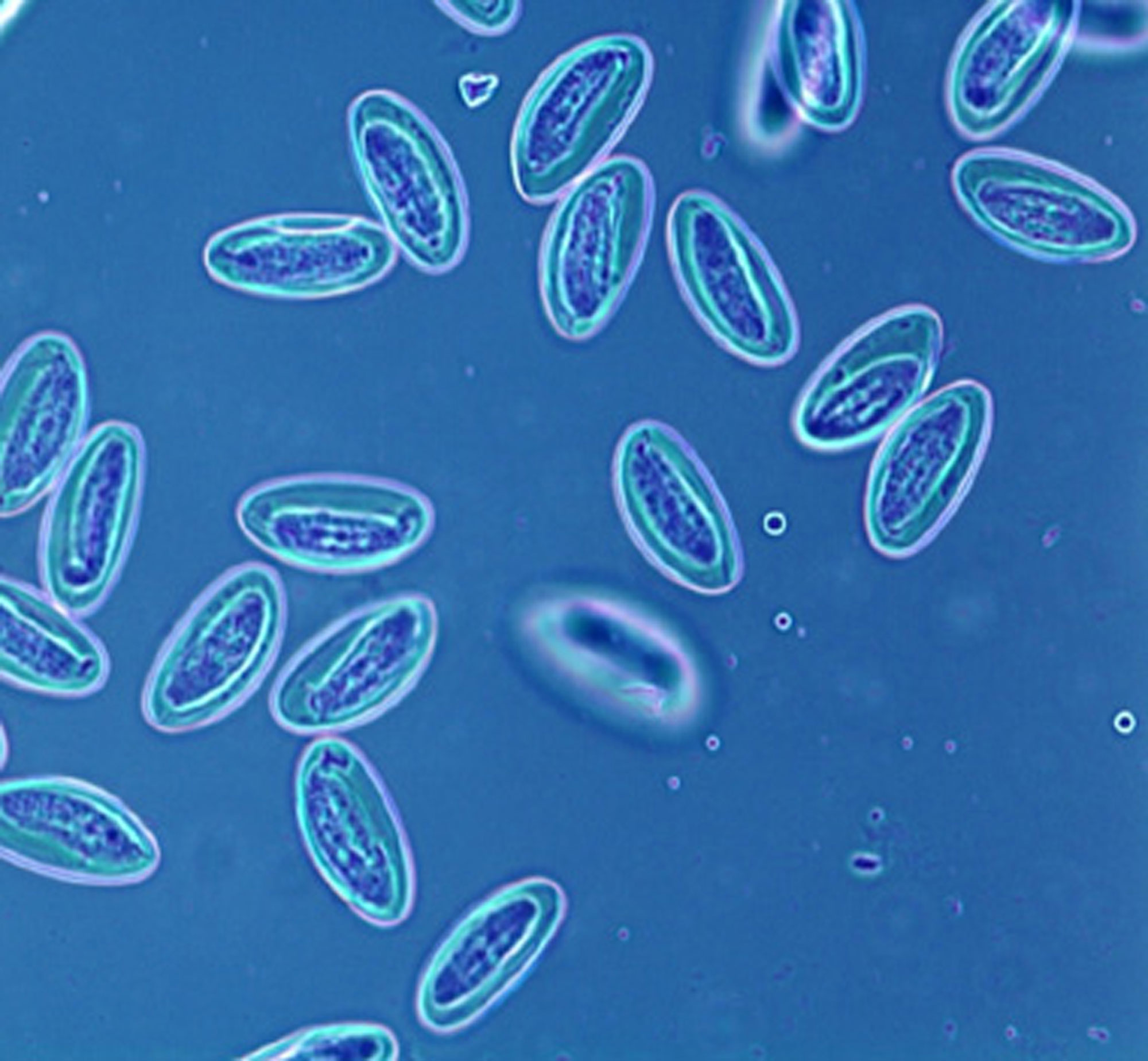
| 2 september 2011 kl. 14.59 | EnterobiusÄggML-a.jpg (fil) |  |
310 kbyte | Foto: Marianne Lebbad, Smittskyddsinstitutet | 1 |
| 2 september 2011 kl. 14.54 | E.histolytTrofAM.jpg (fil) |  |
512 kbyte | Foto: Anders Magnusson, Smittskyddsinstitutet | 1 |
| 2 september 2011 kl. 14.50 | E.histoldisparCystaML-a.jpg (fil) |  |
151 kbyte | Foto: Marianne Lebbad, Smittskyddsinstitutet | 1 |
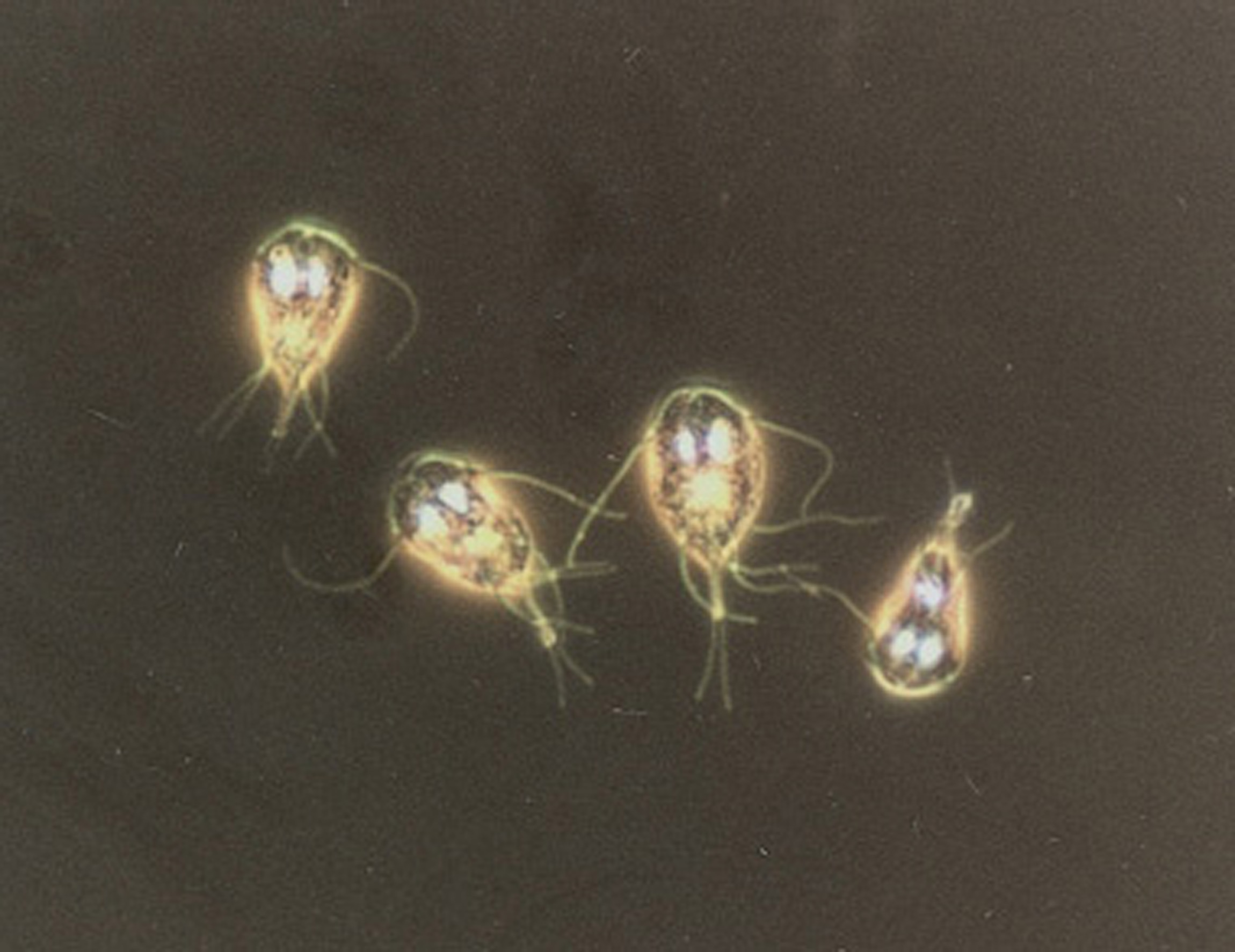
| 2 september 2011 kl. 13.35 | GiardiaTrofozAM.jpg (fil) |  |
334 kbyte | Foto: Anders Magnusson, Smittskyddsinstitutet | 1 |
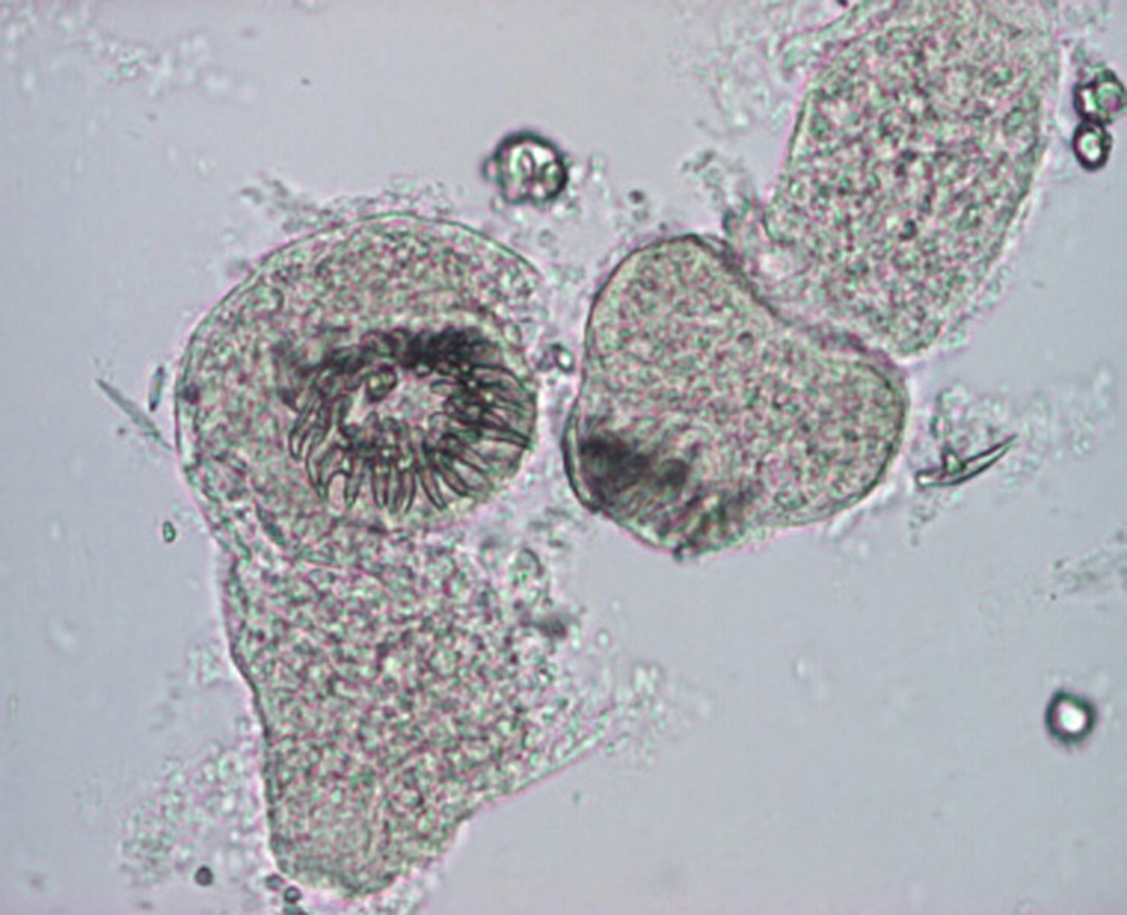
| 2 september 2011 kl. 13.31 | EchinoccProtoscolicesML.jpg (fil) |  |
586 kbyte | Foto: Marianne Lebbad, Smittskyddsinstitutet | 1 |
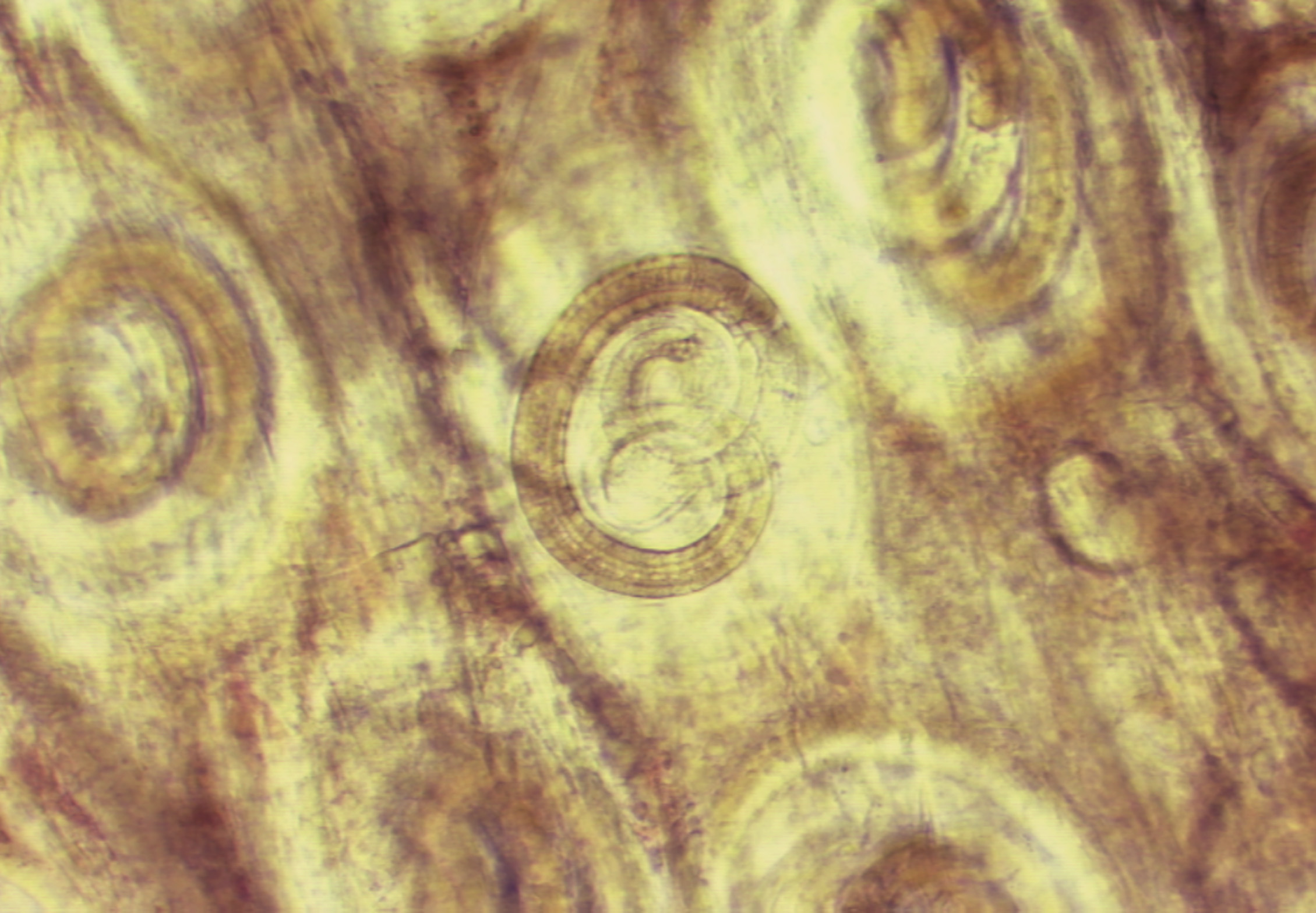
| 2 september 2011 kl. 13.21 | TrichinellaMuskelcystorSB.jpg (fil) |  |
566 kbyte | Foto: Silvia Botero Kleiven, Smittskyddsinstitutet | 1 |
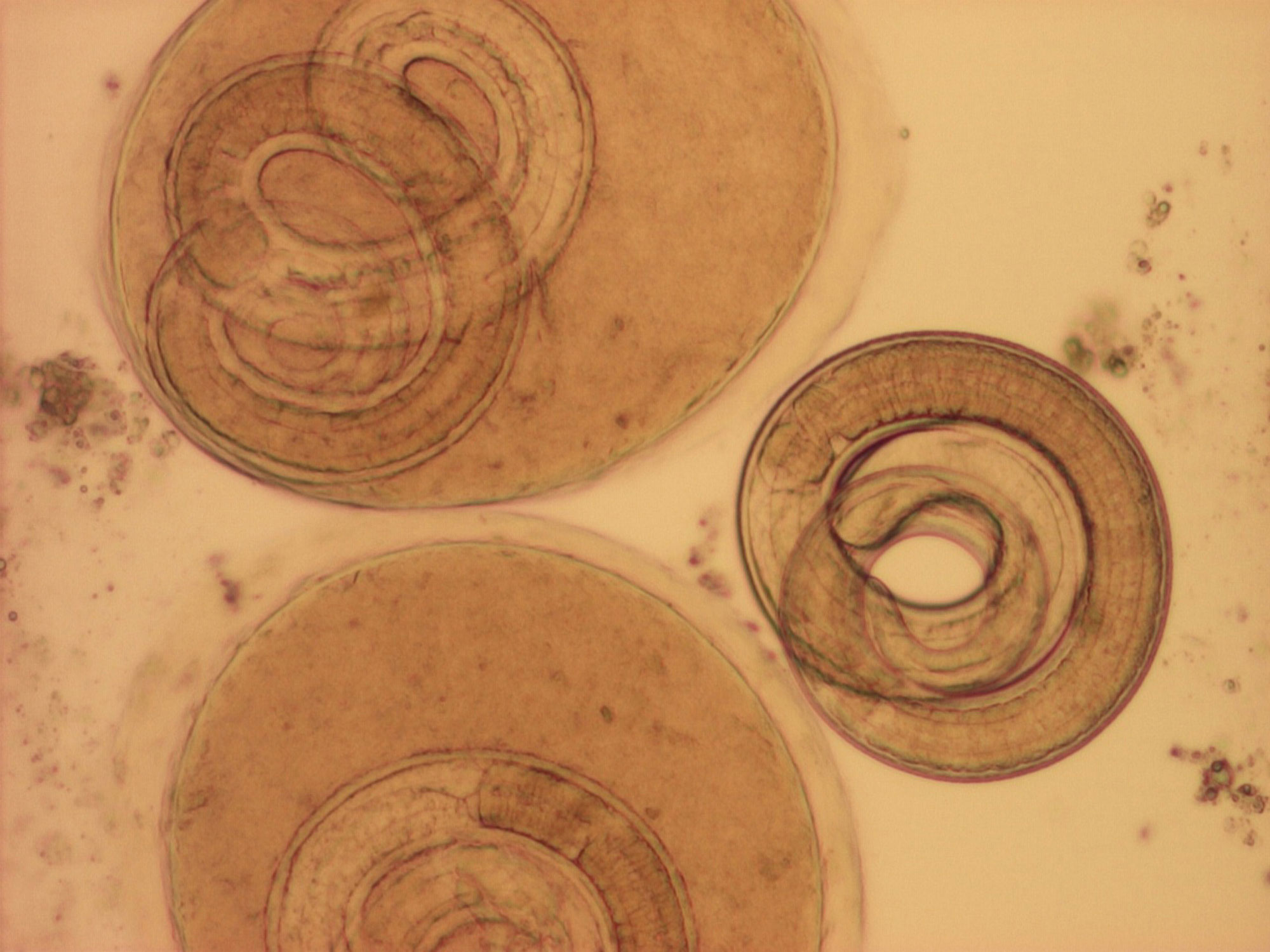
| 2 september 2011 kl. 13.11 | TrichinellaLarverJL-a.jpg (fil) |  |
315 kbyte | Foto: Johan Lindh, Smittskyddsinstitutet | 1 |
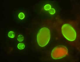
| 2 september 2011 kl. 12.19 | Oocysts of C. parvum (upper left) and cysts of Giardia intestinalis (lower right) IFA.jpg (fil) | _and_cysts_of_Giardia_intestinalis_(lower_right)_IFA.jpg) |
4 kbyte | Foto: Wikimedia commons | 1 |
| 29 augusti 2011 kl. 13.39 | Ac troph..jpg (fil) |  |
53 kbyte | Foto: Jadwiga Winiecka-Krusnell | 1 |
| 29 augusti 2011 kl. 13.23 | Acanth cysts.jpg (fil) |  |
1,82 Mbyte | Foto: Jadwiga Winiecka-Krusnell | 1 |
| 29 augusti 2011 kl. 13.12 | Ac.troph..jpg (fil) |  |
28 kbyte | Foto: Jadwiga Winiecka-Krusnell | 1 |
| 26 augusti 2011 kl. 15.45 | LeishmAmastigML.jpg (fil) |  |
275 kbyte | Foto: Marianne Lebbad, Smittskyddsinstitut | 1 |
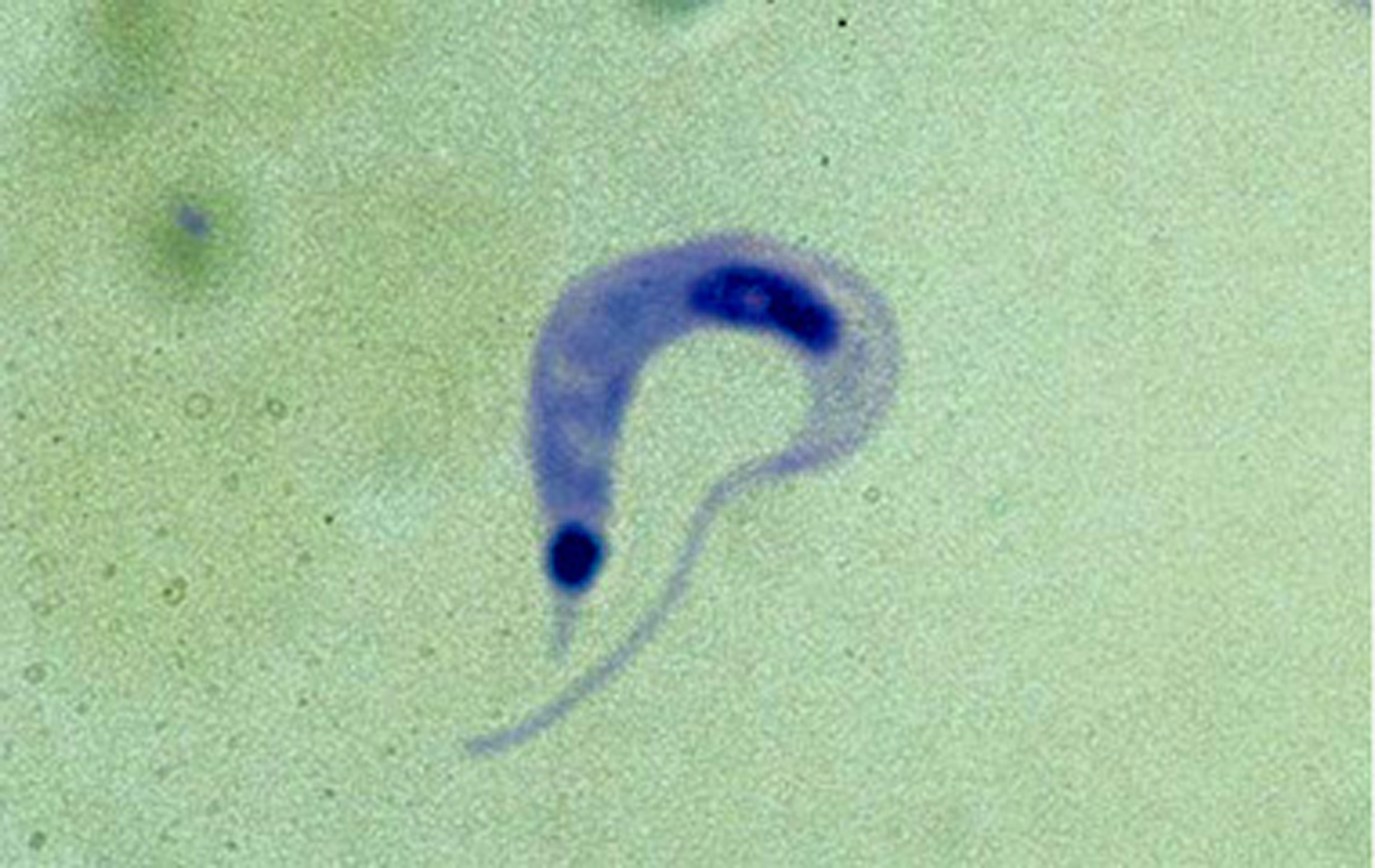
| 26 augusti 2011 kl. 15.35 | TrypbruceiBS-a.jpg (fil) |  |
129 kbyte | Foto: Silvia Botero, Smittskyddsinstitutet | 1 |
| 26 augusti 2011 kl. 15.21 | TrypcruziAM.jpg (fil) |  |
397 kbyte | Foto: Anders Magnusson, Smittskyddsinstitutet | 1 |
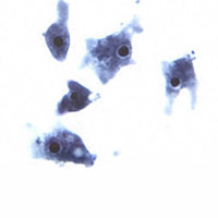
| 26 augusti 2011 kl. 13.23 | AcanthamoebaTrofJWK.jpg (fil) |  |
206 kbyte | Foto: Jadwiga Winiecka-Krusnell | 1 |
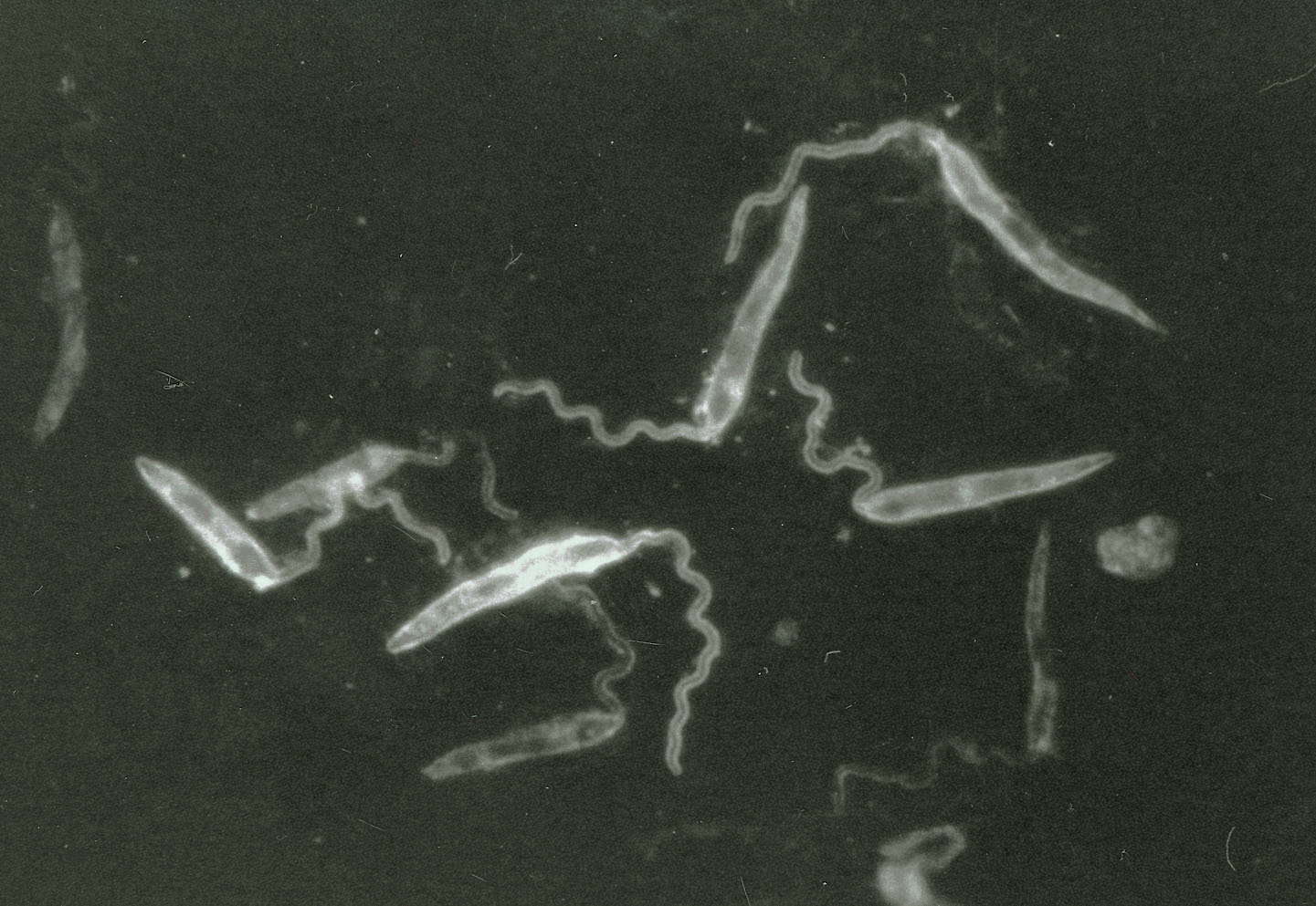
| 26 augusti 2011 kl. 12.38 | Leishmaniapromastigotes.jpg (fil) |  |
318 kbyte | Foto; Jadwiga Winiecka-Krusnell, Smittskyddsinstitutet | 1 |
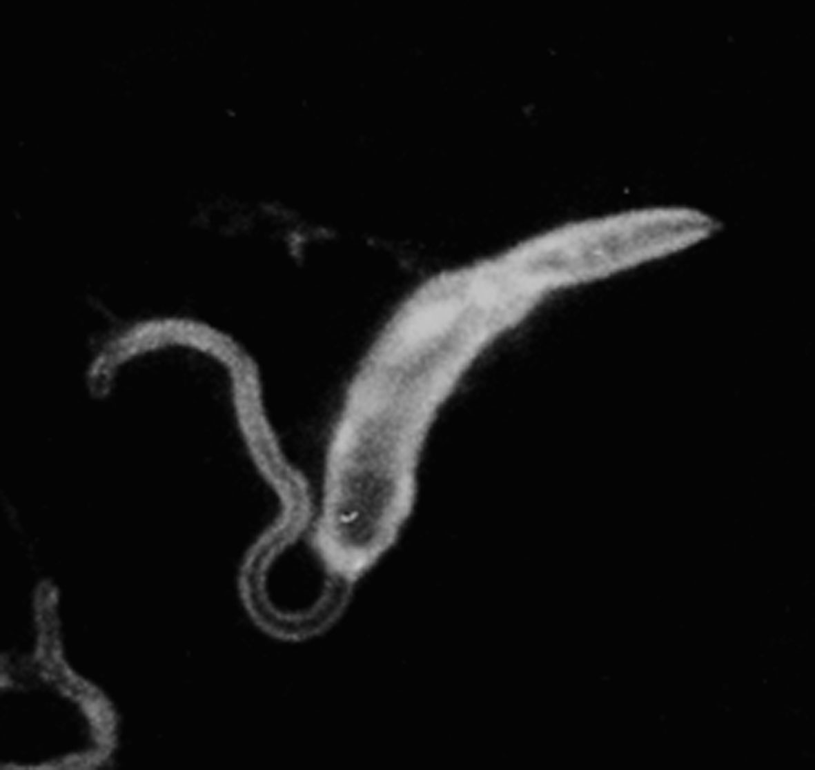
| 26 augusti 2011 kl. 12.22 | Leishmaniapromastigote.jpg (fil) |  |
61 kbyte | Foto: Jadwiga Winiecka-Krusnell, Smittskyddsinstitutet | 1 |
| 26 augusti 2011 kl. 10.26 | T. cruzi amastigotes.crop..png (fil) |  |
1,4 Mbyte | Foto: Jadwiga Winiecka-Krusnell | 1 |
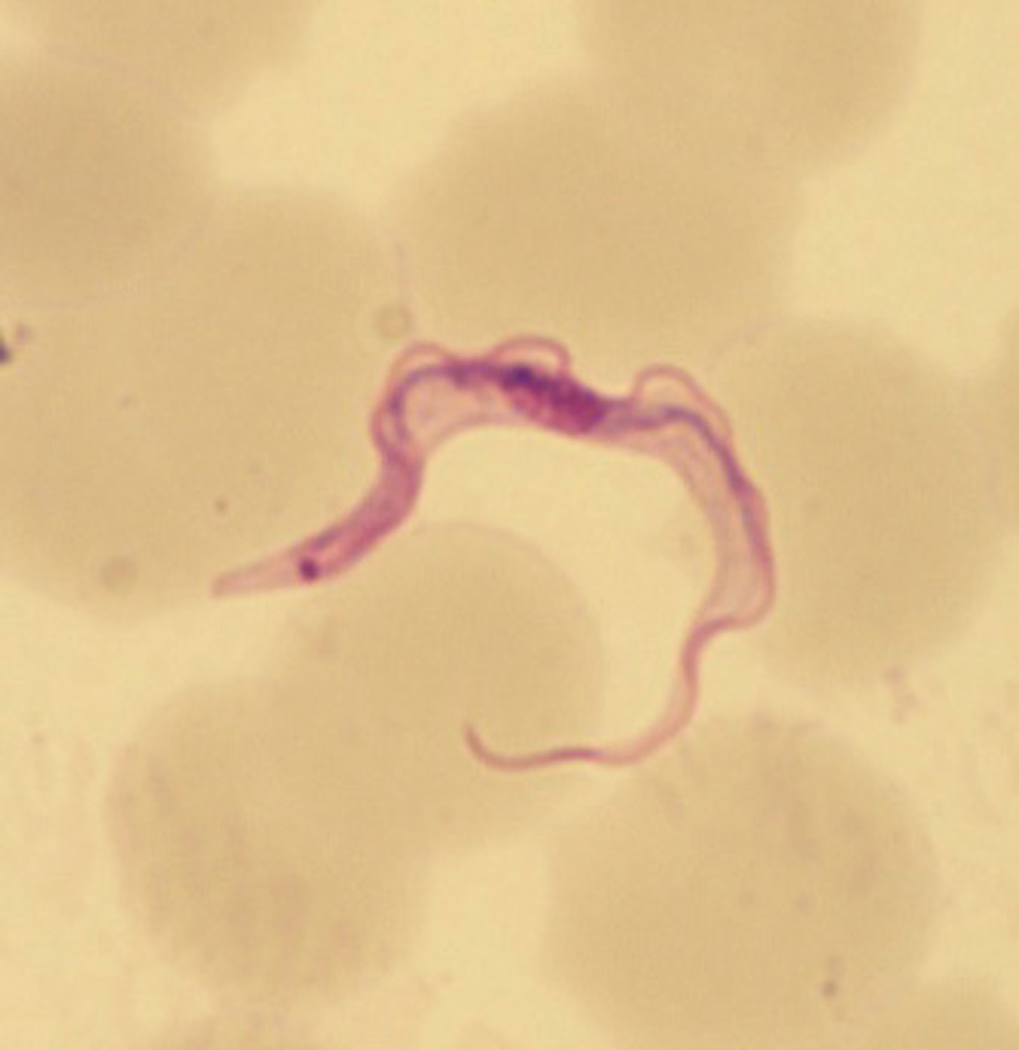
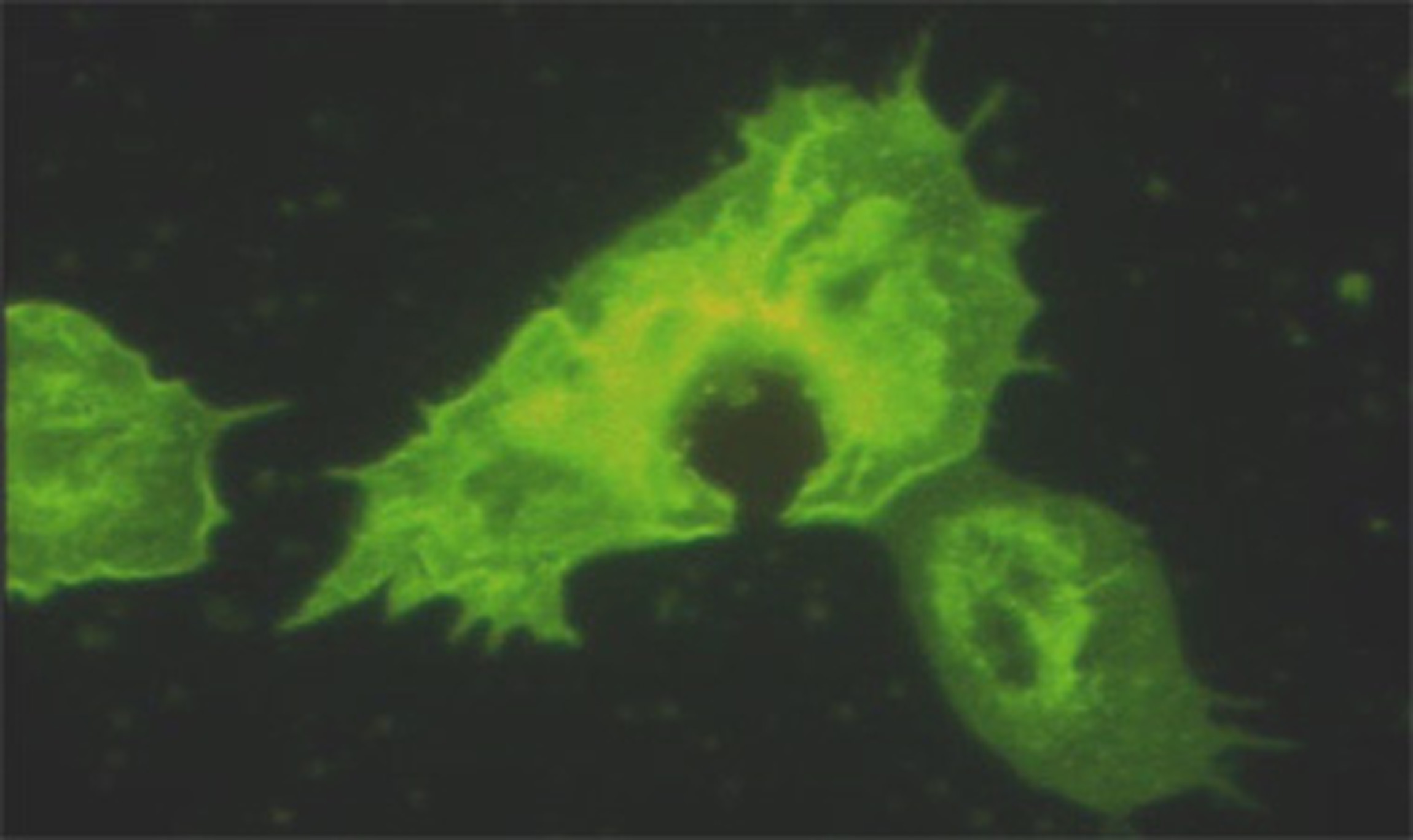
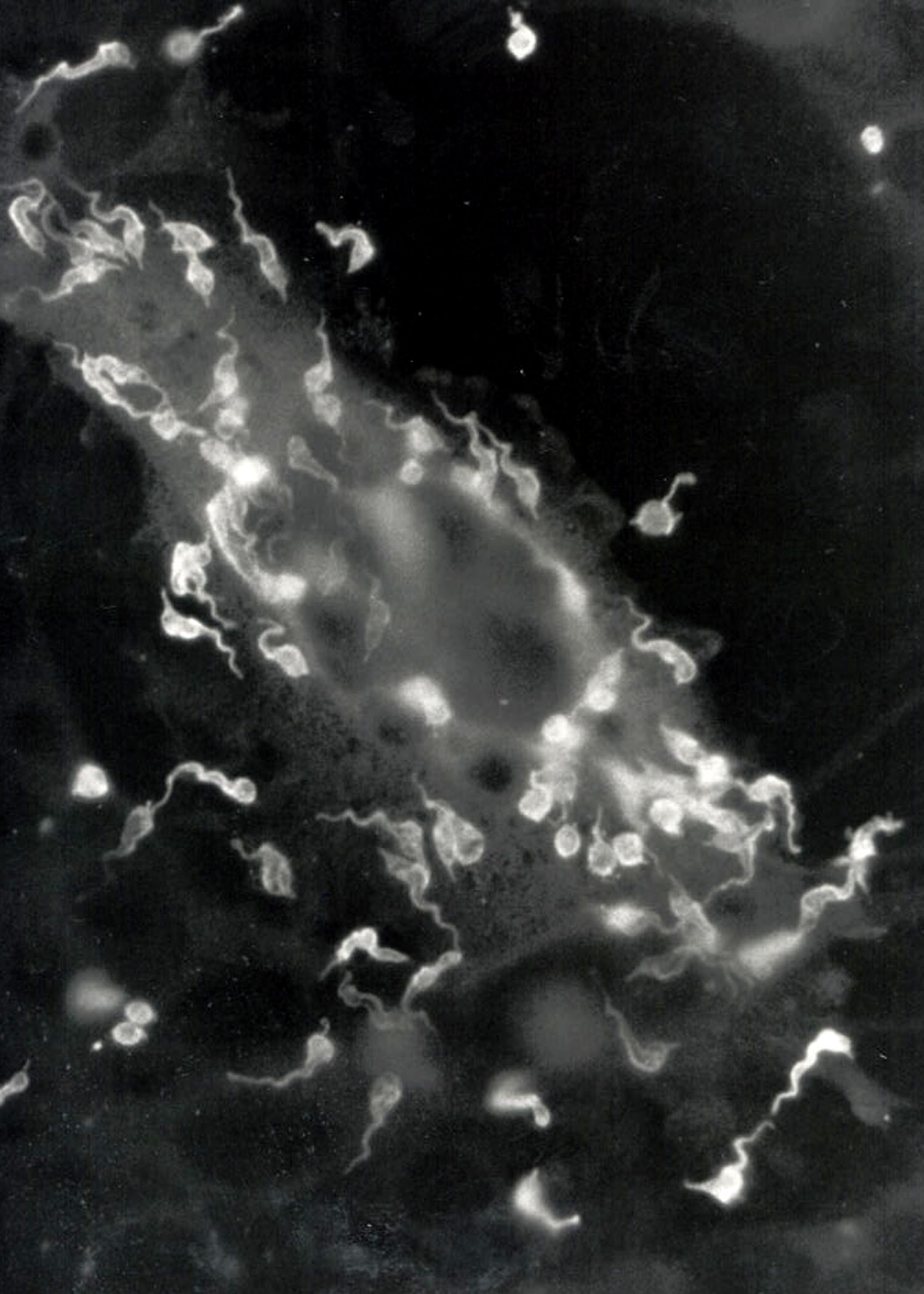
| 26 augusti 2011 kl. 10.09 | T.cruzi trypomastigotes 2.jpg (fil) |  |
297 kbyte | T. cruzi trypomastigoter från in vitro odling på glioma-celler. foto: Jadwiga Winiecka-Krusnell, Smittskyddsinstitutet | 1 |
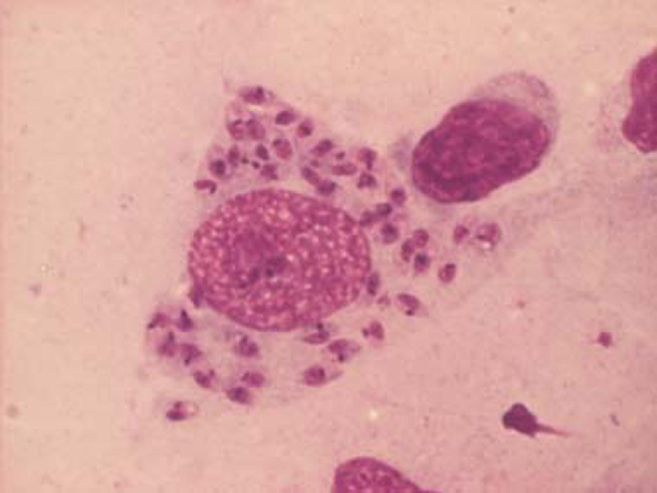
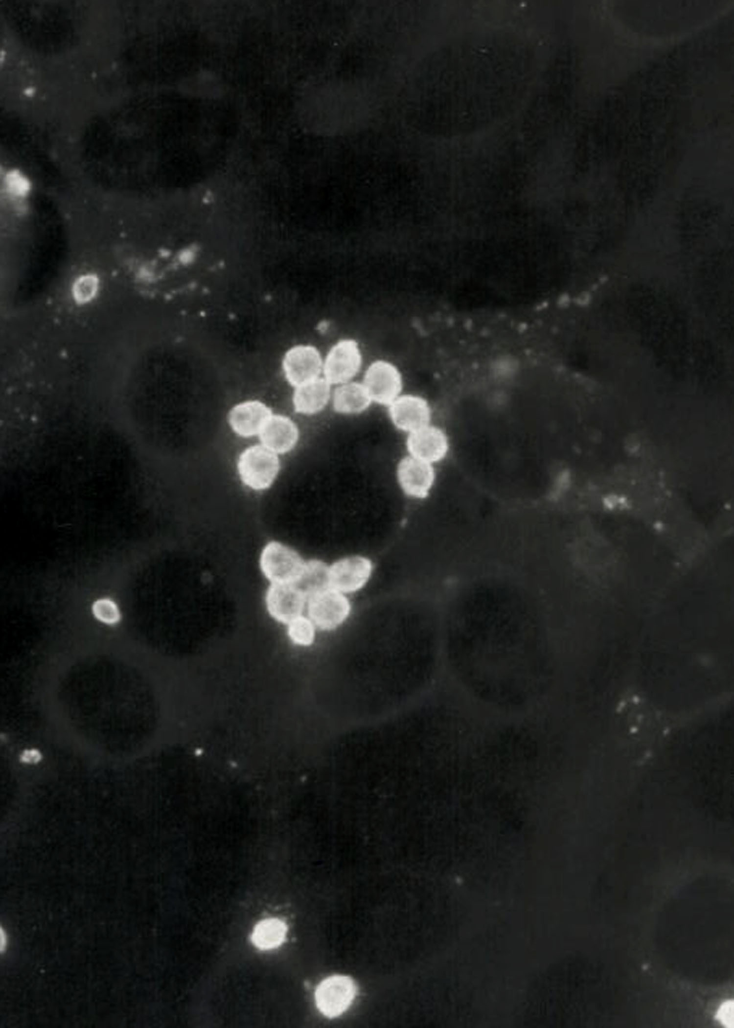
| 26 augusti 2011 kl. 10.06 | T. cruzi amastigotes.crop..jpg (fil) |  |
204 kbyte | T. cruzi amastigoter från in vitro odling på glioma-celler. Foto: Jadwiga Winiecka-Krusnell, Smittskyddsinstitutet | 1 |
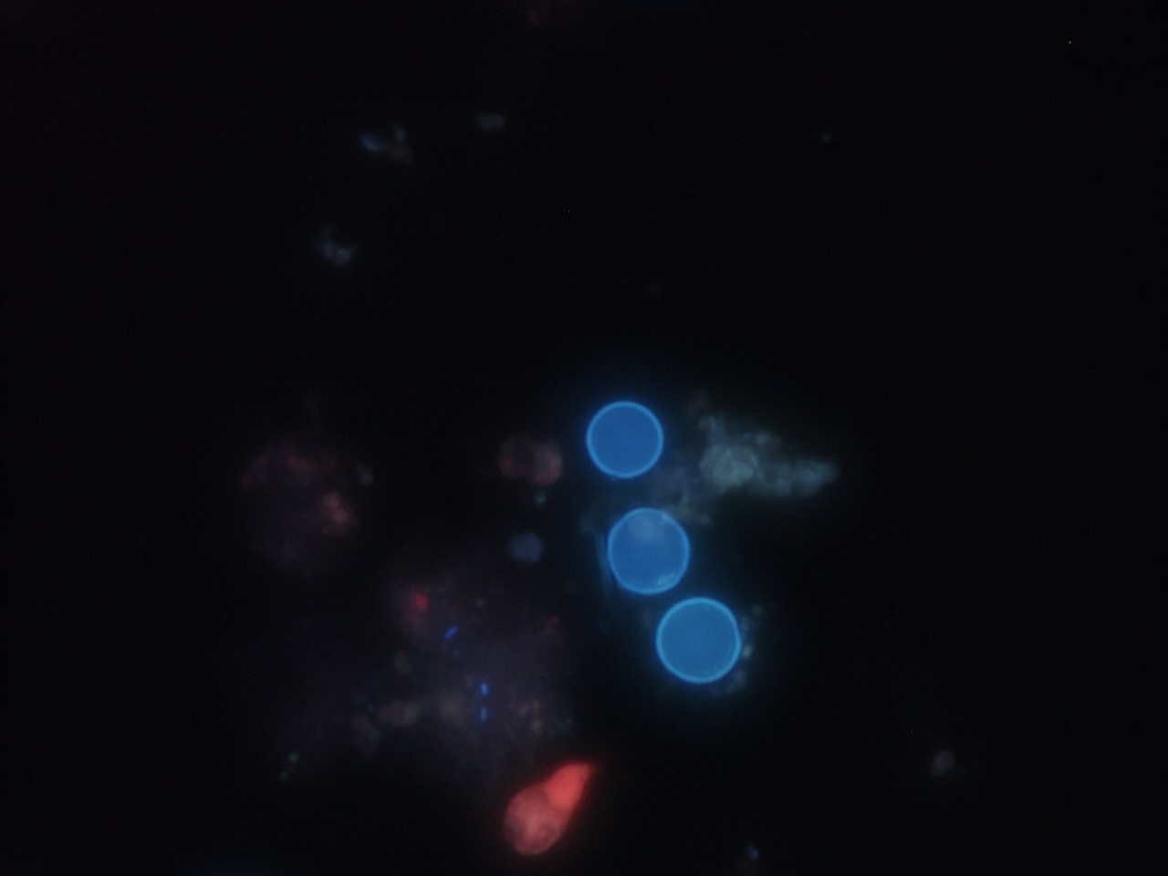
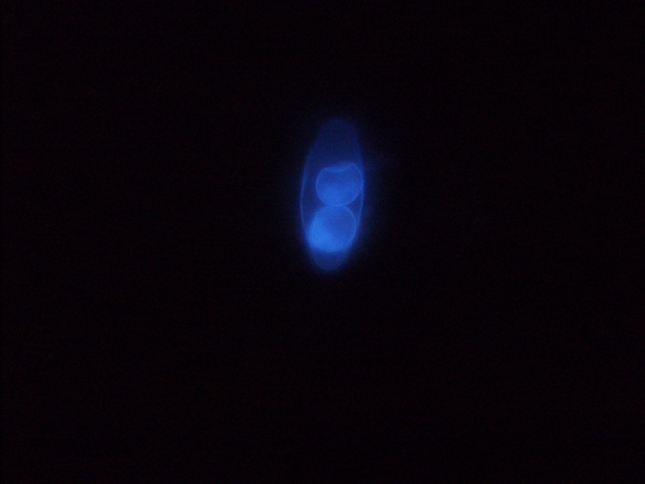
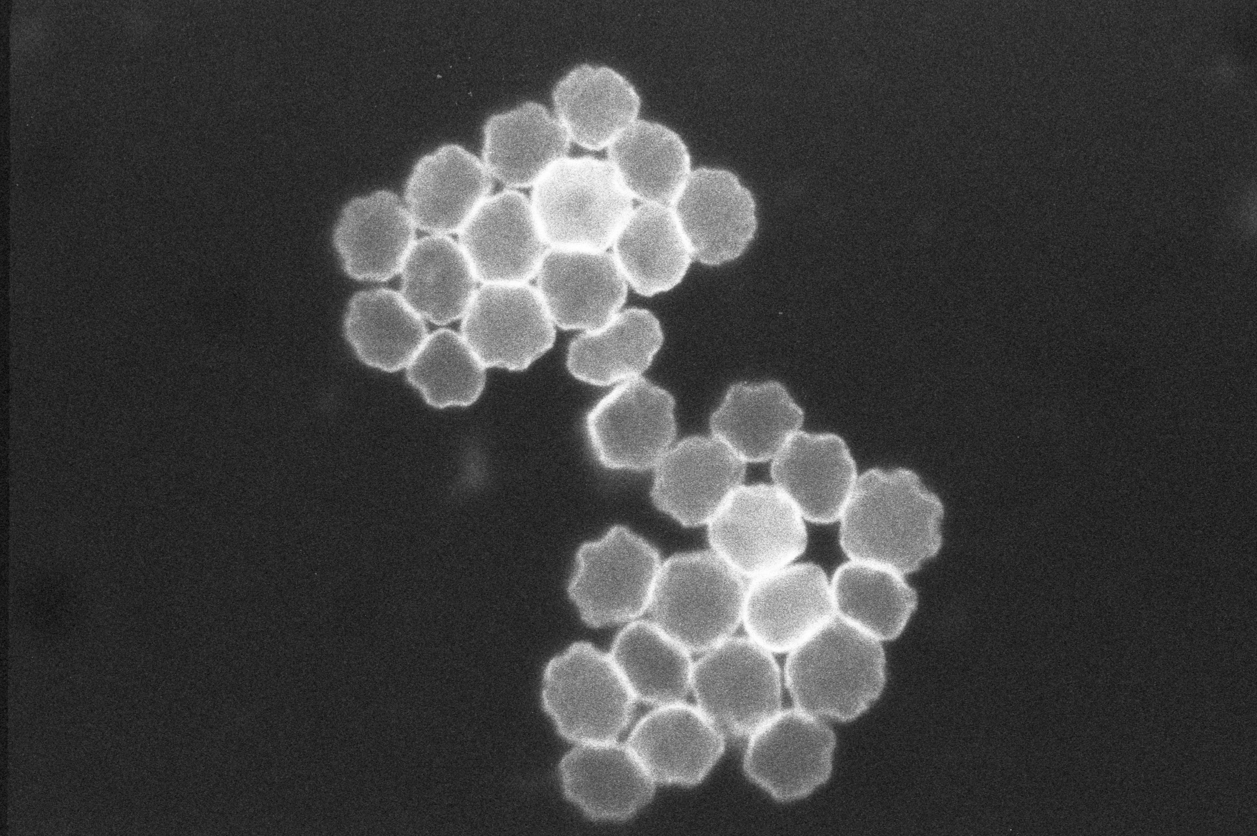
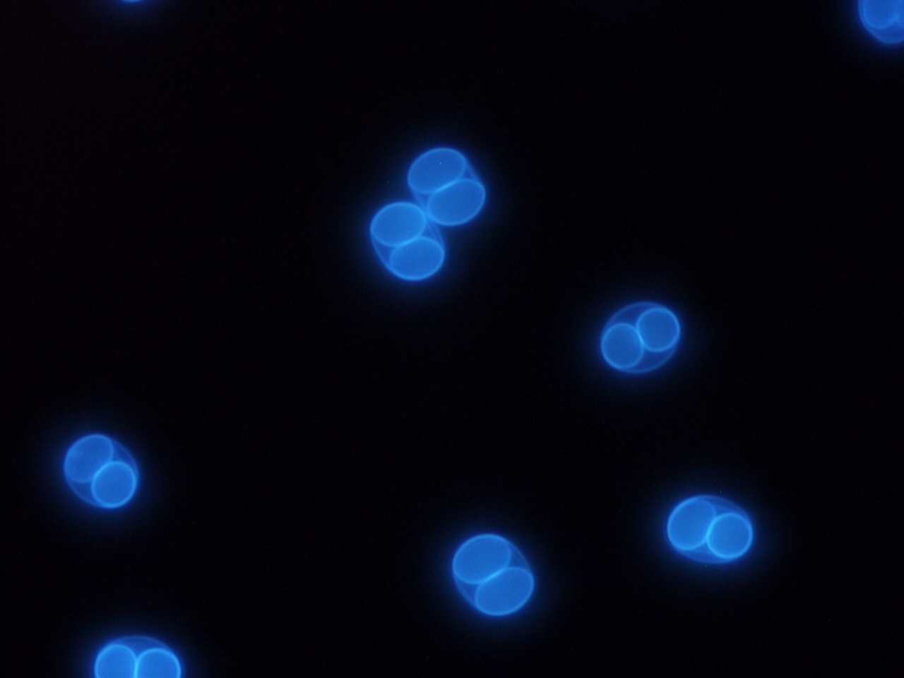
| 26 augusti 2011 kl. 09.16 | Jadwiga Toxo oocysts.jpg (fil) |  |
590 kbyte | Autofluorescerande oocystor av T. gondi | 1 |
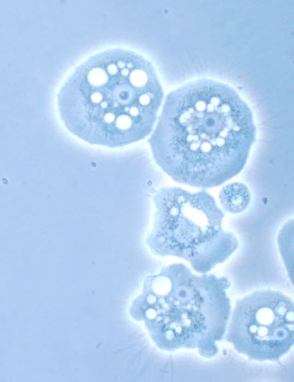
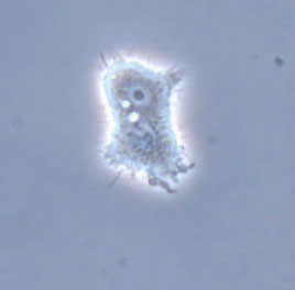
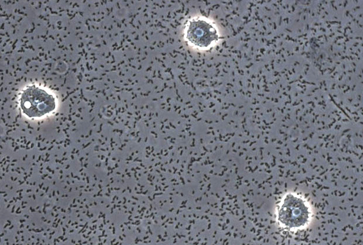
| 19 augusti 2011 kl. 11.59 | Jadwiga Acanthamoeba 40 obj. .0006.jpg (fil) |  |
207 kbyte | Acanthamoeba. Foto Jadwiga Krusnell | 1 |
| 19 augusti 2011 kl. 11.55 | Leishmania1.jpg (fil) |  |
319 kbyte | Leishmania promastigotes i RPMI. Foto Jadwiga Krusnell | 1 |
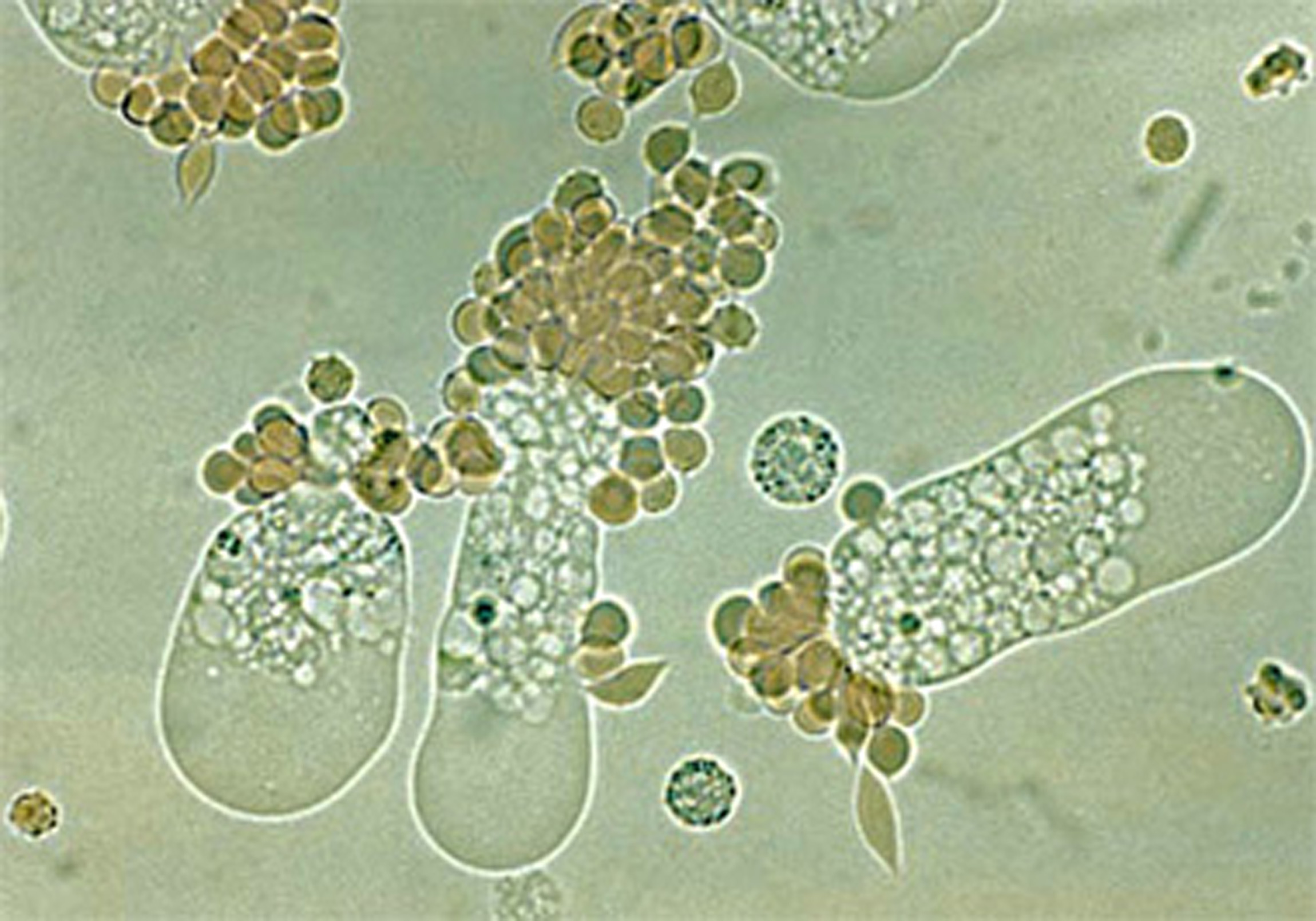
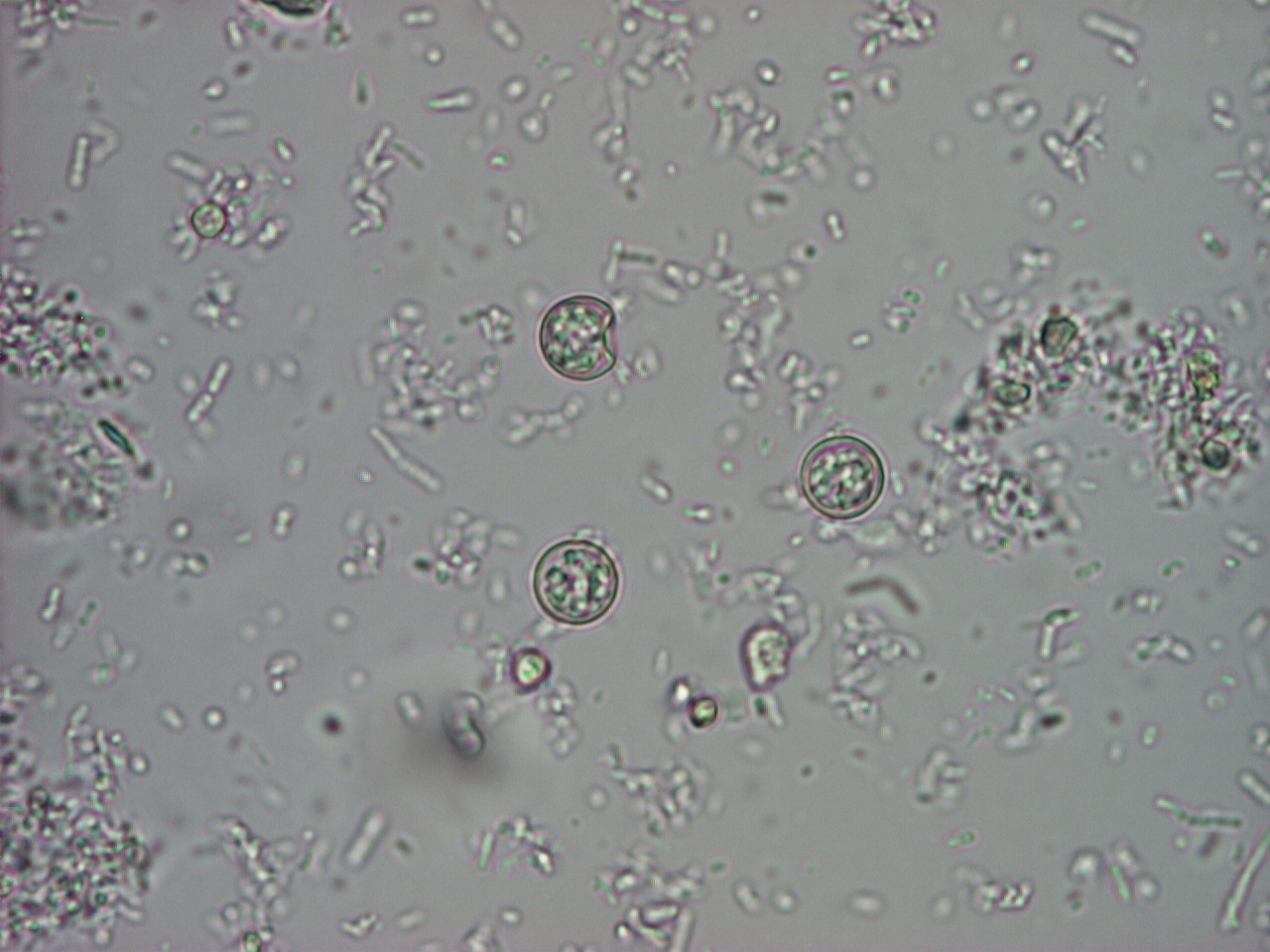
| 19 augusti 2011 kl. 11.49 | Cyclospora2 100x.jpg (fil) |  |
233 kbyte | Cyklospora Bild 2. Foto Marianne Lebbad | 1 |
| 19 augusti 2011 kl. 11.45 | Cyklospora 1.jpg (fil) |  |
290 kbyte | Cyclospora Bild 1. Foto: Marianne Lebbad | 1 |
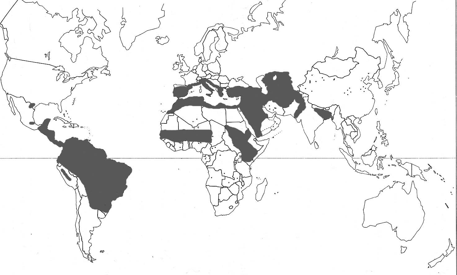
| 19 augusti 2011 kl. 11.36 | Leishmaniakarta 3.jpg (fil) |  |
120 kbyte | 1 | |
| 19 augusti 2011 kl. 11.30 | Leishmania promastigotes.jpg (fil) |  |
308 kbyte | Leishmania_promastigotes i RPMI-medium. Foto Jadwiga Krusnell | 1 |