Uppladdningar av Jadwiga Krusnell
Hoppa till navigering
Hoppa till sök
Den här specialsidan visar alla filer som laddats upp.
| Datum | Namn | Miniatyrbild | Storlek (byte) | Beskrivning | Versioner |
|---|---|---|---|---|---|
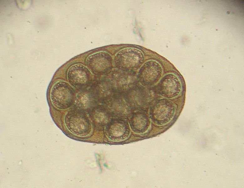
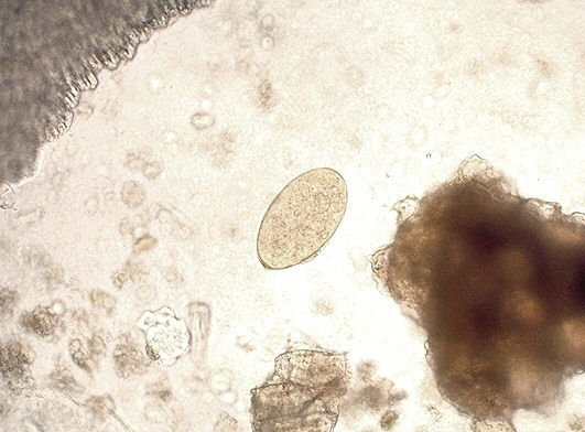
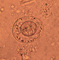
| 14 september 2011 kl. 14.19 | 779px-Dipylidium caninum ovum 1.jpg (fil) |  |
53 kbyte | Foto Joel Mills via Wikimedia Commons | 2 |
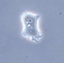
| 29 augusti 2011 kl. 13.12 | Ac.troph..jpg (fil) |  |
28 kbyte | Foto: Jadwiga Winiecka-Krusnell | 1 |
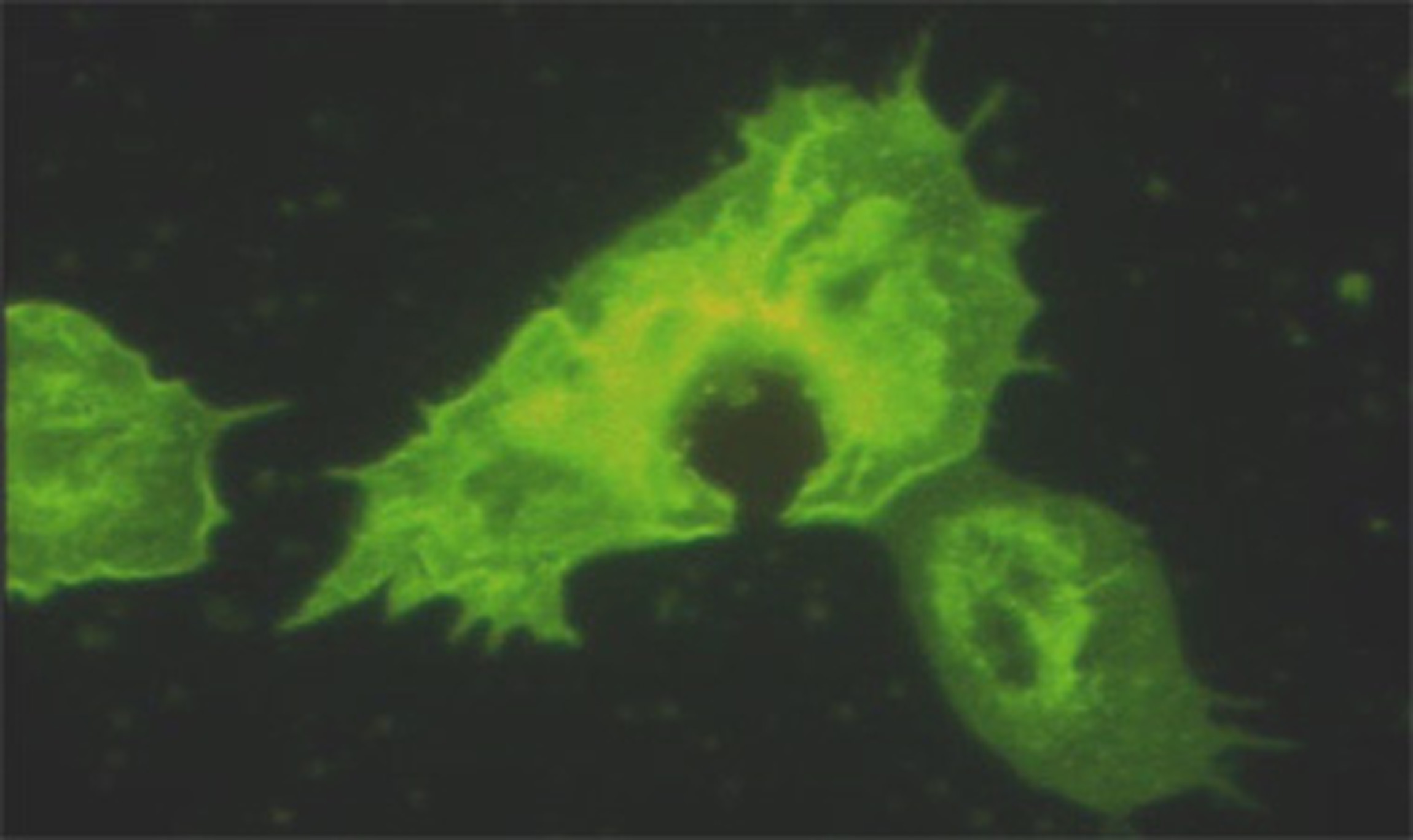
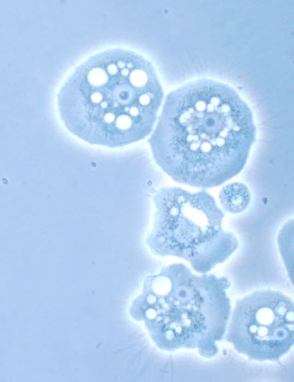
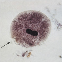
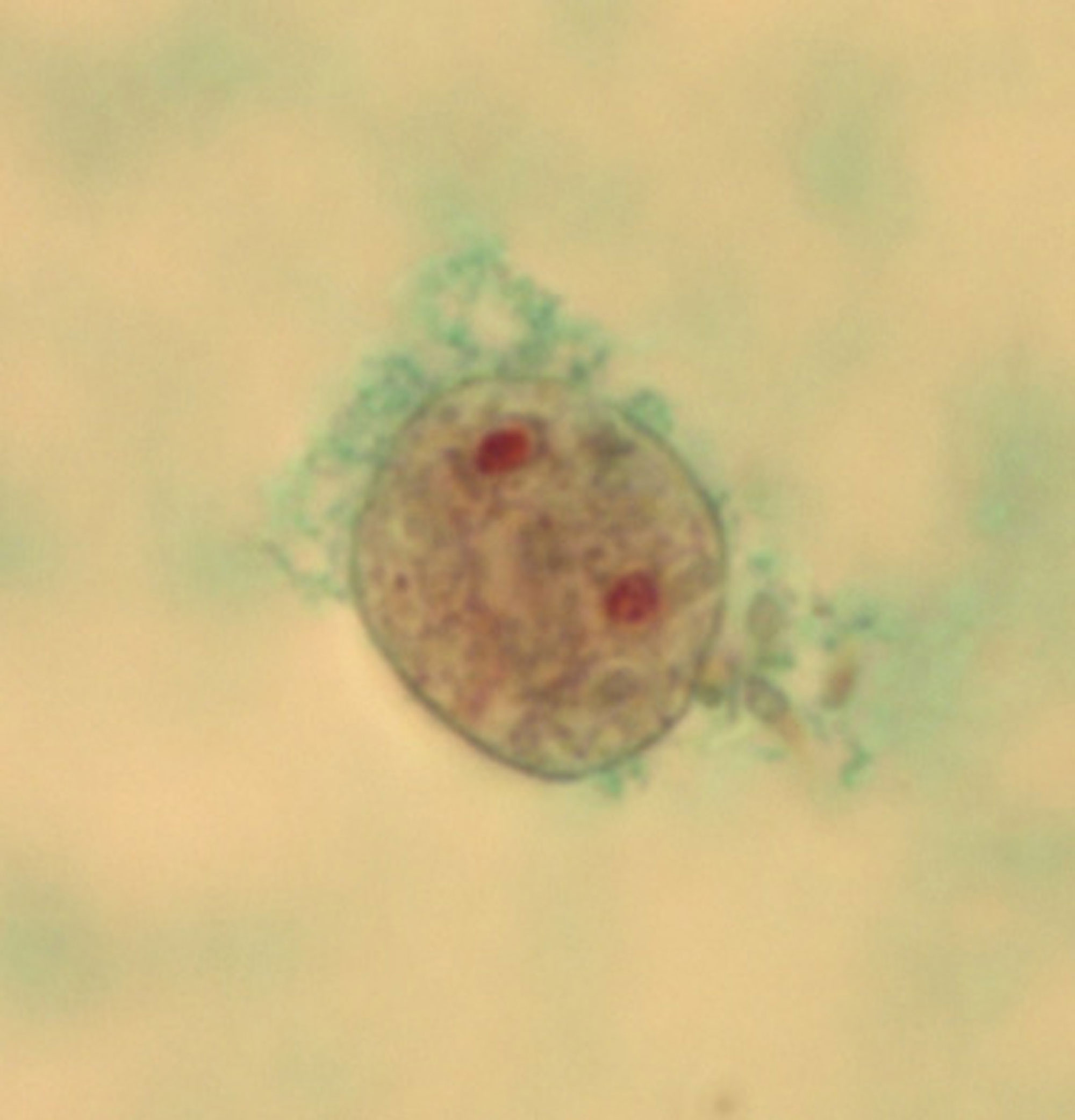
| 26 augusti 2011 kl. 13.23 | AcanthamoebaTrofJWK.jpg (fil) |  |
206 kbyte | Foto: Jadwiga Winiecka-Krusnell | 1 |
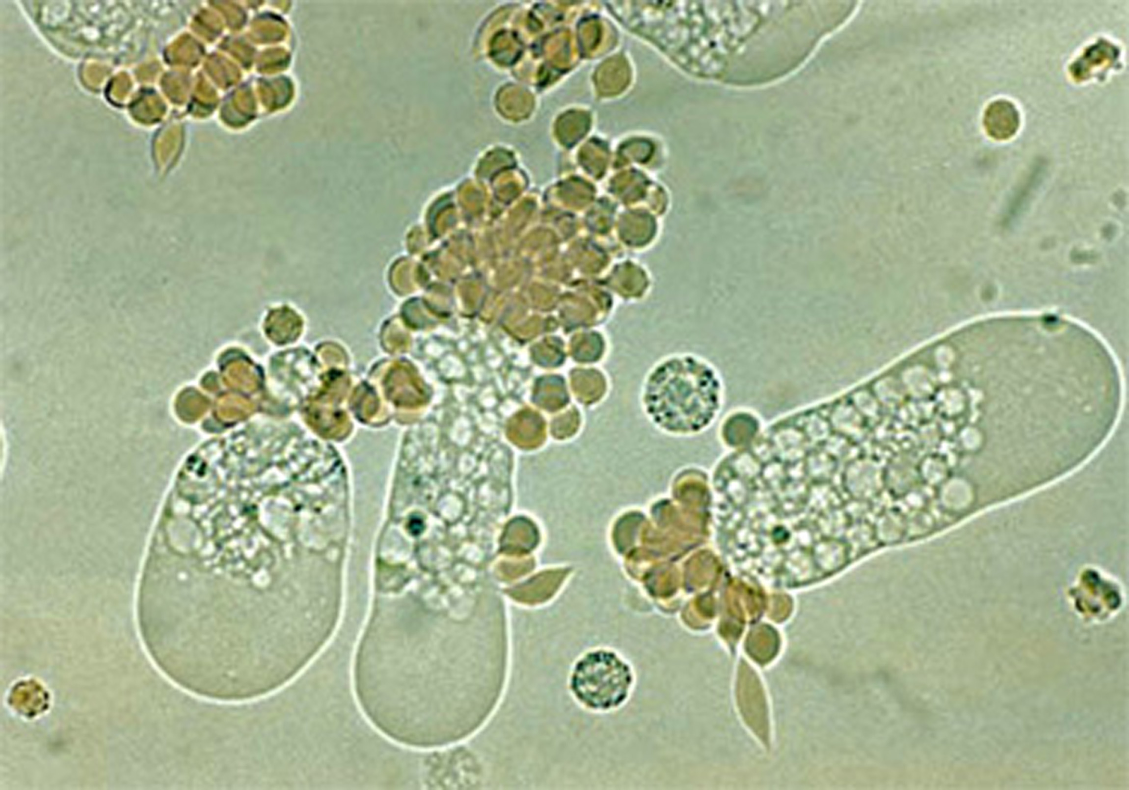
| 29 augusti 2011 kl. 13.23 | Acanth cysts.jpg (fil) |  |
1,82 Mbyte | Foto: Jadwiga Winiecka-Krusnell | 1 |
| 29 augusti 2011 kl. 13.39 | Ac troph..jpg (fil) |  |
53 kbyte | Foto: Jadwiga Winiecka-Krusnell | 1 |
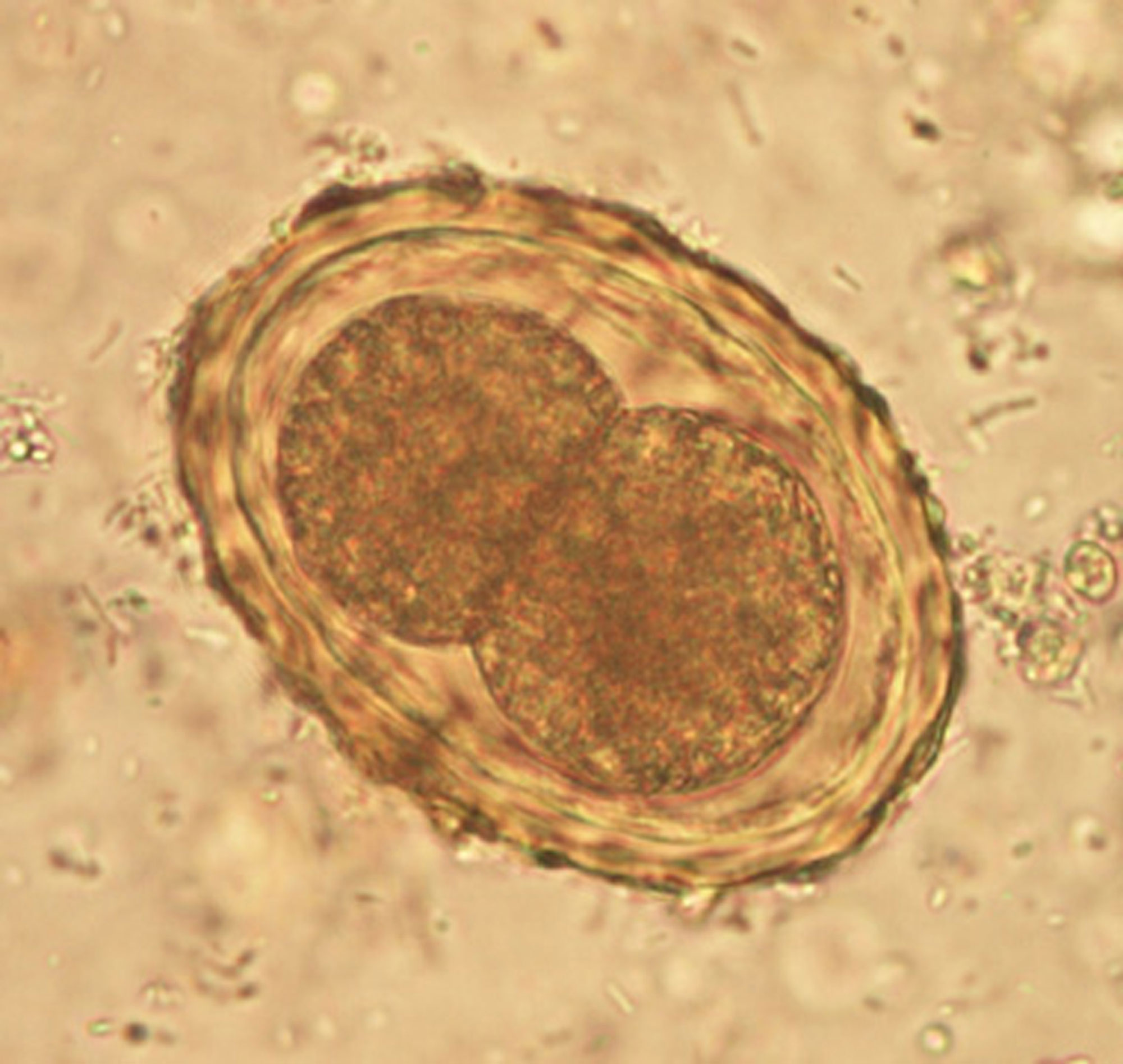
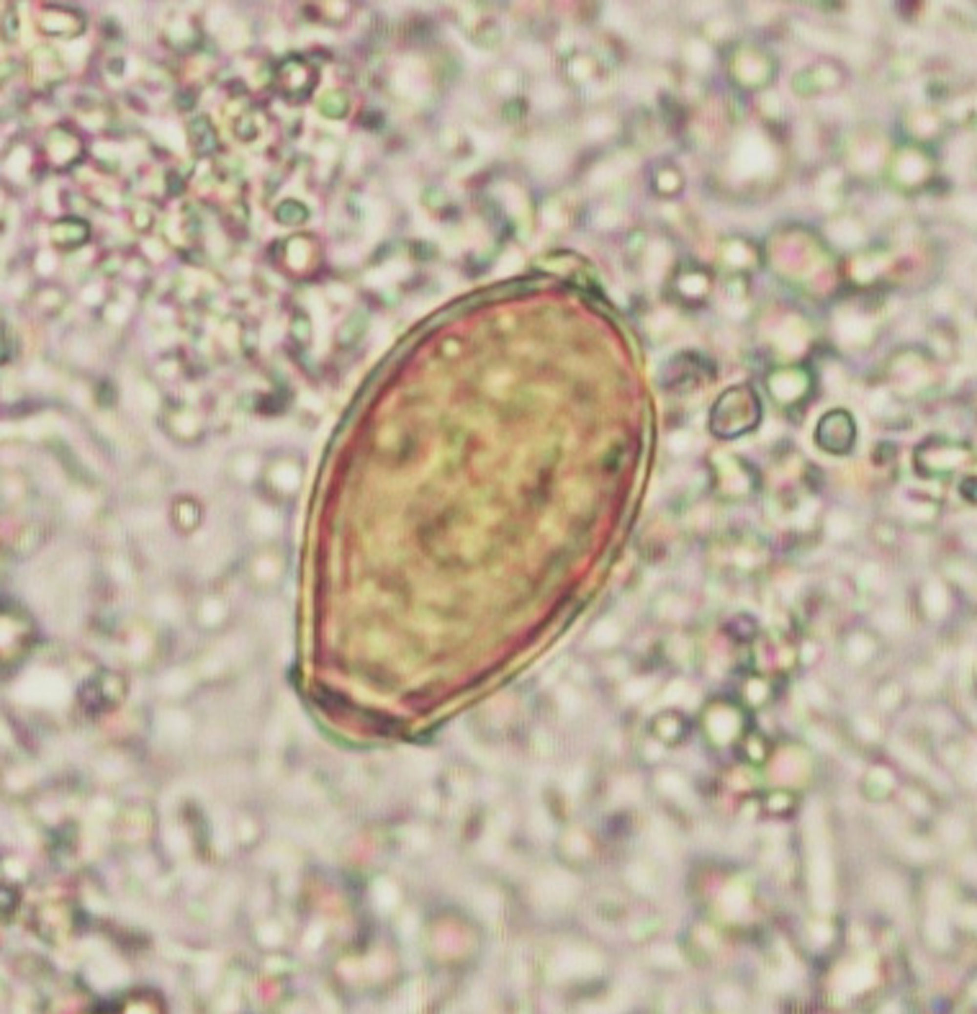
| 28 mars 2012 kl. 10.39 | AscarisÄggMLa.jpg (fil) |  |
299 kbyte | Foto: Marianne Lebbad, SMI | 1 |
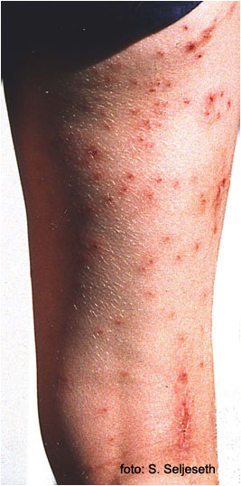
| 26 mars 2012 kl. 15.55 | Badklåda ben.jpg (fil) |  |
87 kbyte | Utseende:se symtom. Foto: Stein Seljeseth | 1 |
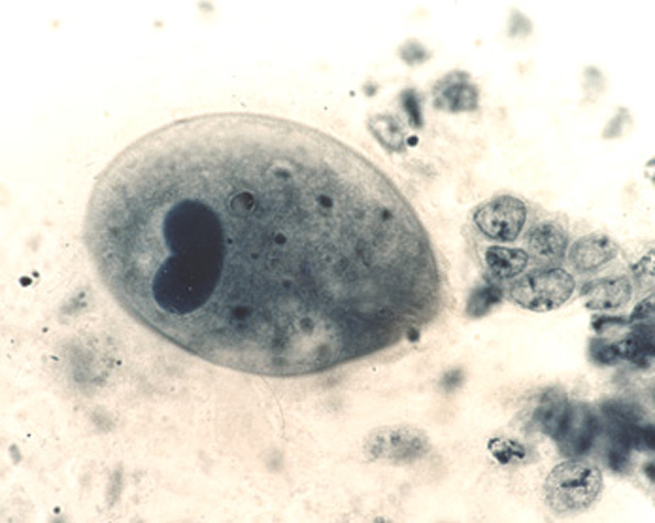
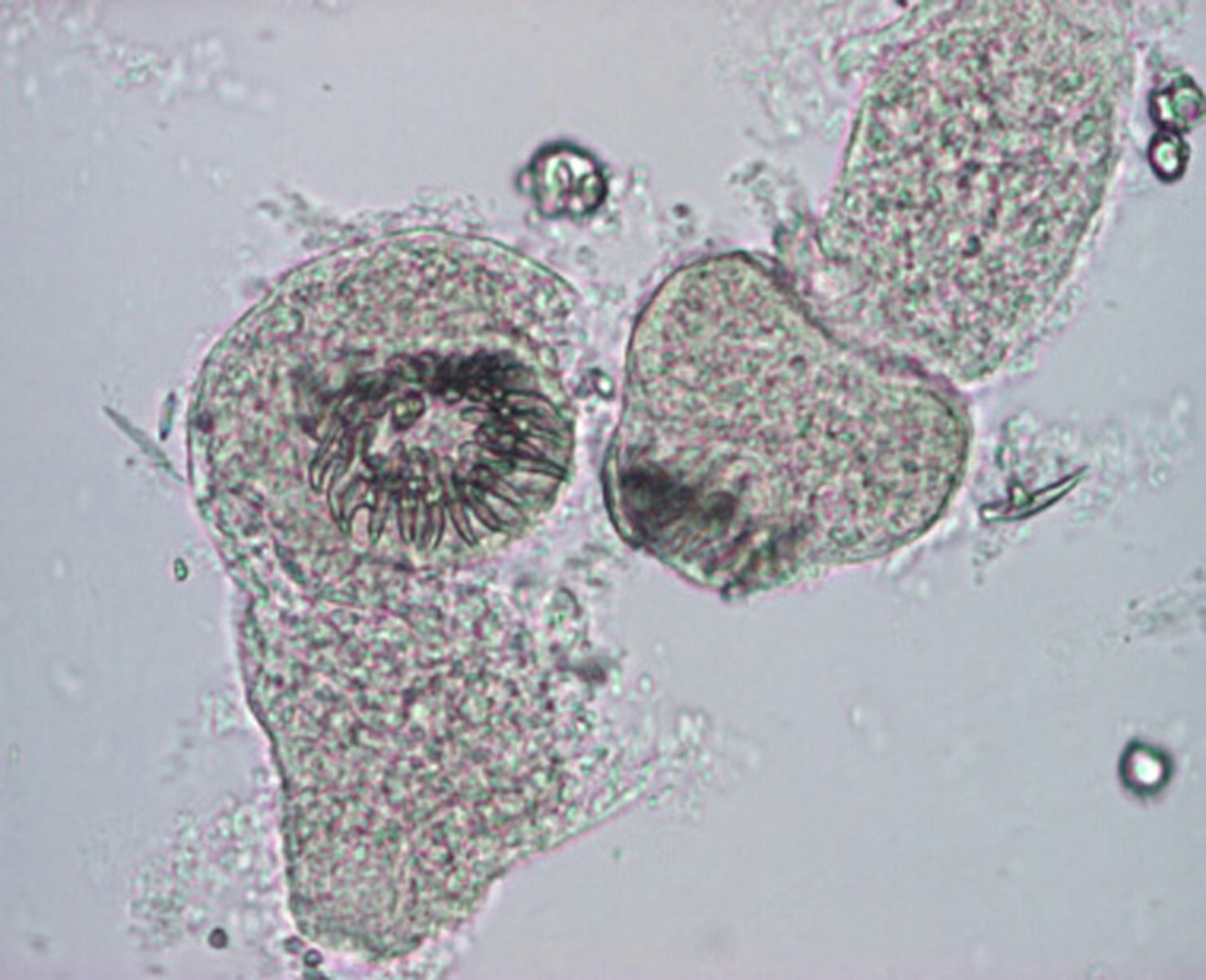
| 28 mars 2012 kl. 10.42 | BalantidiumTrofAM.jpg (fil) |  |
369 kbyte | Foto: Anders Magnusson, SMI | 1 |
| 14 mars 2012 kl. 17.12 | Balantidium coli wet mount.jpg (fil) |  |
305 kbyte | från Wiki media commons | 1 |
| 14 mars 2012 kl. 17.13 | Balantidium trophB.jpg (fil) |  |
12 kbyte | från wiki media commons | 1 |
| 28 mars 2012 kl. 10.42 | BlastocystisML-a.jpg (fil) |  |
126 kbyte | Foto: Marianne Lebbad, SMI | 1 |
| 7 september 2011 kl. 12.20 | Clonorchis sinensis egg 06G0049 jpg lores.jpg (fil) |  |
51 kbyte | Foto: CDC via Wikimedia Commons | 1 |
| 7 september 2011 kl. 10.19 | ClonorchOpisthÄggBS-a.jpg (fil) |  |
344 kbyte | Foto: Silvia Botero Kleiven, Smittskyddsinstitutet | 1 |
| 15 augusti 2012 kl. 15.17 | Cryptosporidium Ziehl Alae Gati.jpg (fil) |  |
42 kbyte | Ocystor av Cryptosporidium spp, Z-N, bild från Wiki media | 1 |
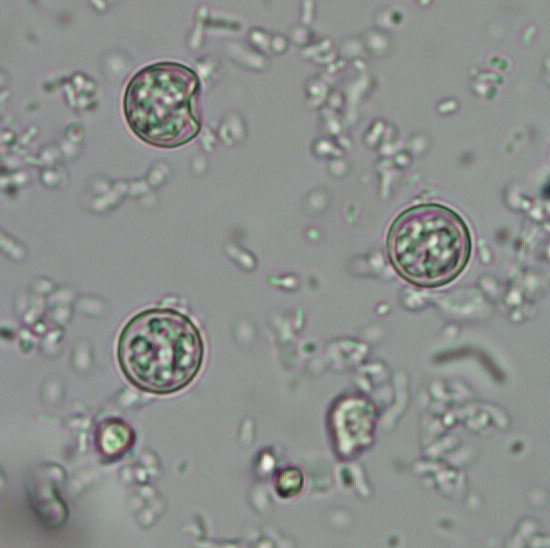
| 19 augusti 2011 kl. 11.49 | Cyclospora2 100x.jpg (fil) |  |
233 kbyte | Cyklospora Bild 2. Foto Marianne Lebbad | 1 |
| 14 mars 2012 kl. 15.47 | Cyclospora Bild 2. 100x.jpg (fil) |  |
233 kbyte | 1 | |
| 6 mars 2012 kl. 16.27 | Cyclospora bild 3.jpeg (fil) |  |
157 kbyte | 1 | |
| 14 mars 2012 kl. 15.48 | Cyclospora bild 3 crop.jpg (fil) |  |
39 kbyte | 1 | |
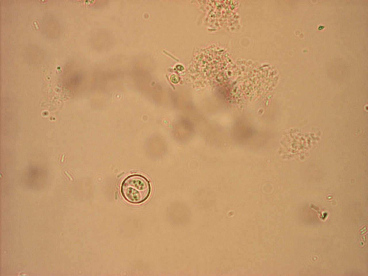
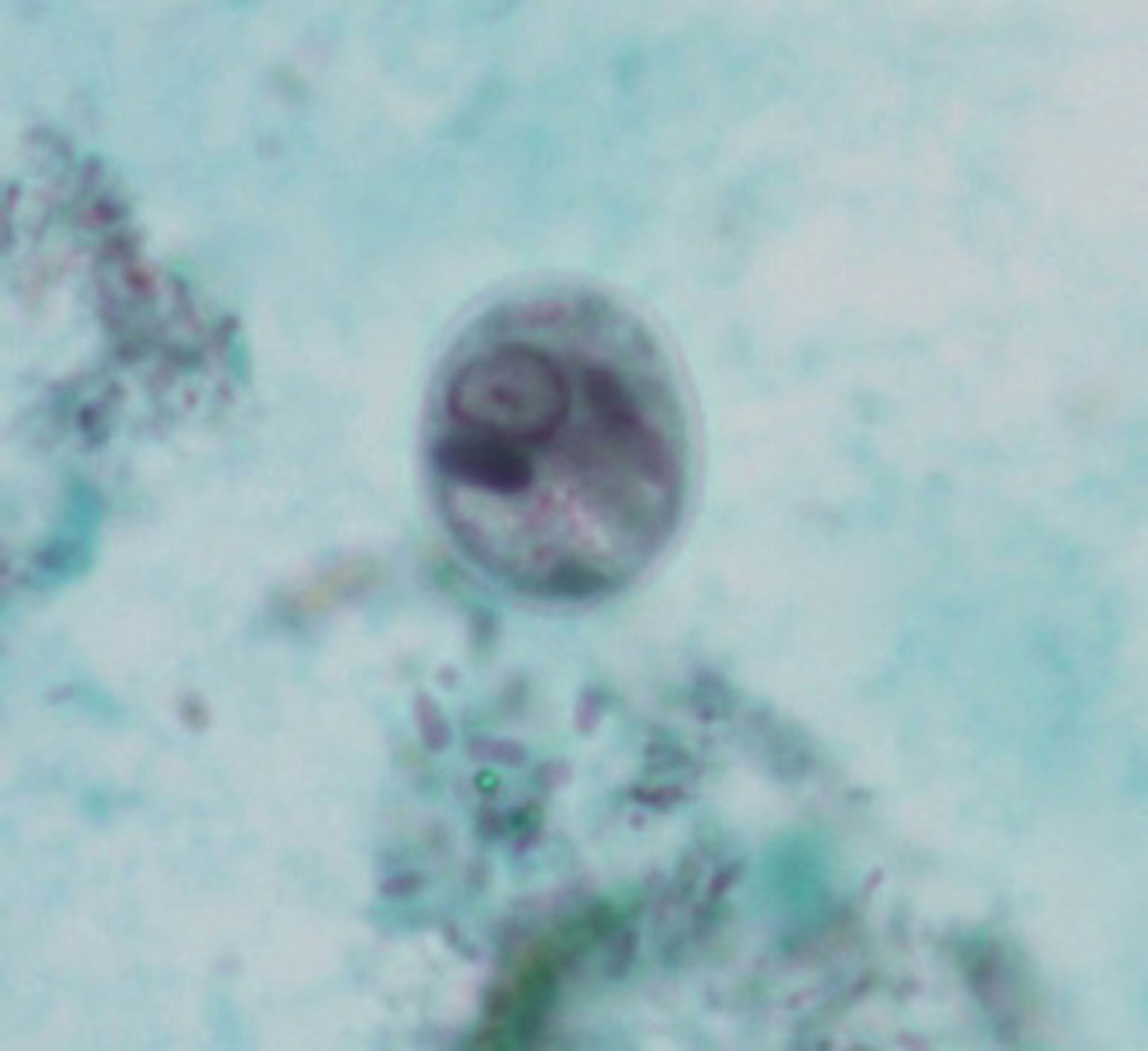
| 6 mars 2012 kl. 16.29 | Cyclospora Bild 4 sporulerad oocysta.jpeg (fil) |  |
226 kbyte | 1 | |
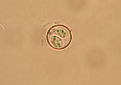
| 6 mars 2012 kl. 16.34 | Cyclospora Bild 4 sporulerad oocysta crop.jpg (fil) |  |
36 kbyte | 1 | |
| 14 mars 2012 kl. 15.43 | Cyclospora crop Bild 1.jpg (fil) |  |
69 kbyte | 1 | |
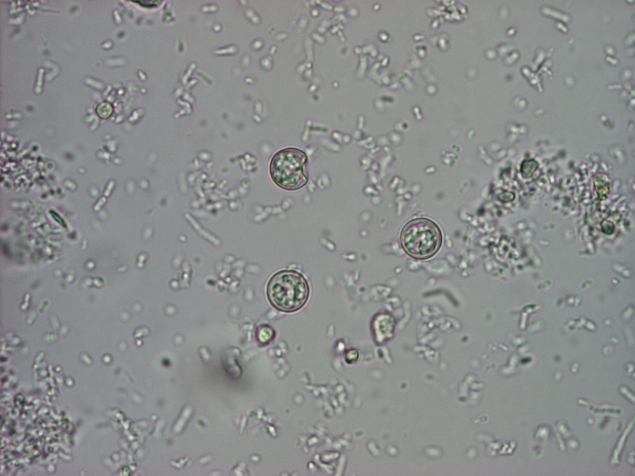
| 19 augusti 2011 kl. 11.45 | Cyklospora 1.jpg (fil) |  |
290 kbyte | Cyclospora Bild 1. Foto: Marianne Lebbad | 1 |
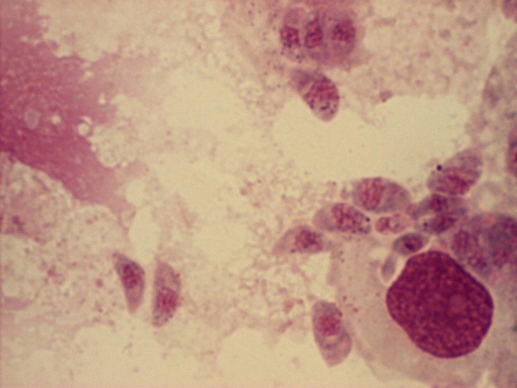
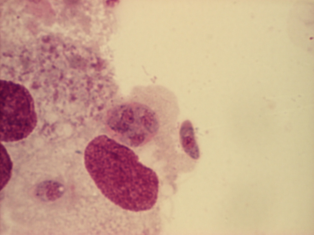
| 19 april 2012 kl. 16.08 | C 100x.jpg (fil) |  |
74 kbyte | Toxoplasma/monocyter, giemsa. Foto Silvia Botero kleiven | 2 |
| 28 mars 2012 kl. 10.43 | DientamoebaTrofM-aL.jpg (fil) |  |
143 kbyte | Foto: Marianne Lebbad, SMI | 1 |
| 8 juni 2012 kl. 14.29 | Dok1.jpg (fil) |  |
45 kbyte | 1 | |
| 6 september 2011 kl. 15.50 | D dendriticum egg wtmt JCG C.jpg (fil) |  |
29 kbyte | Foto: CDC vie Wikimedia Commons | 1 |
| 2 september 2011 kl. 14.50 | E.histoldisparCystaML-a.jpg (fil) |  |
151 kbyte | Foto: Marianne Lebbad, Smittskyddsinstitutet | 1 |
| 2 september 2011 kl. 14.54 | E.histolytTrofAM.jpg (fil) |  |
512 kbyte | Foto: Anders Magnusson, Smittskyddsinstitutet | 1 |
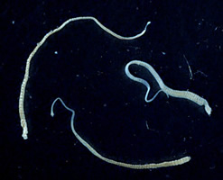
| 2 september 2011 kl. 13.31 | EchinoccProtoscolicesML.jpg (fil) |  |
586 kbyte | Foto: Marianne Lebbad, Smittskyddsinstitutet | 1 |
| 8 september 2011 kl. 15.16 | Egg of Fasciola hepatica 08G0041 lores.jpg (fil) |  |
48 kbyte | Foto: CDC via Wikimedia Commons | 1 |
| 5 september 2011 kl. 14.37 | Ehistdisp cyst wtmt.jpg (fil) |  |
14 kbyte | Foto: Wiki media commons | 1 |
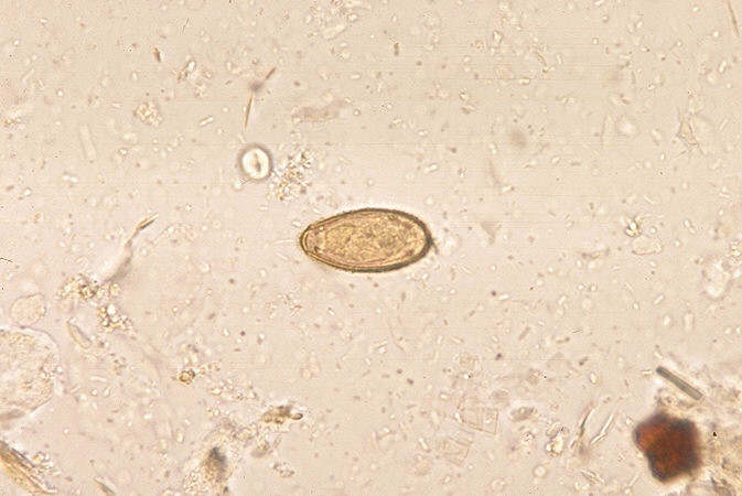
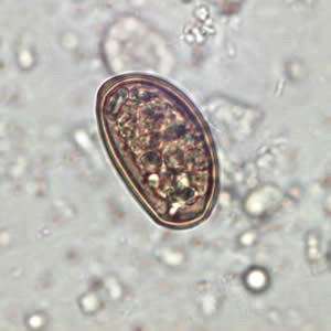
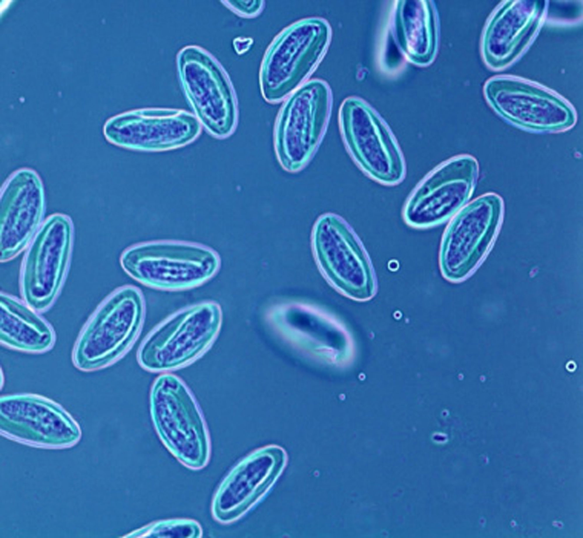
| 2 september 2011 kl. 14.59 | EnterobiusÄggML-a.jpg (fil) |  |
310 kbyte | Foto: Marianne Lebbad, Smittskyddsinstitutet | 1 |
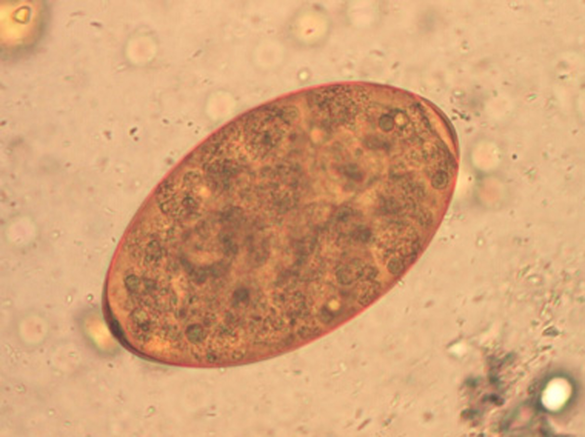
| 8 september 2011 kl. 15.05 | FasciolaÄggML.jpg (fil) |  |
417 kbyte | Foto: Marianne Lebbad, Smittskyddsinstitutet | 1 |
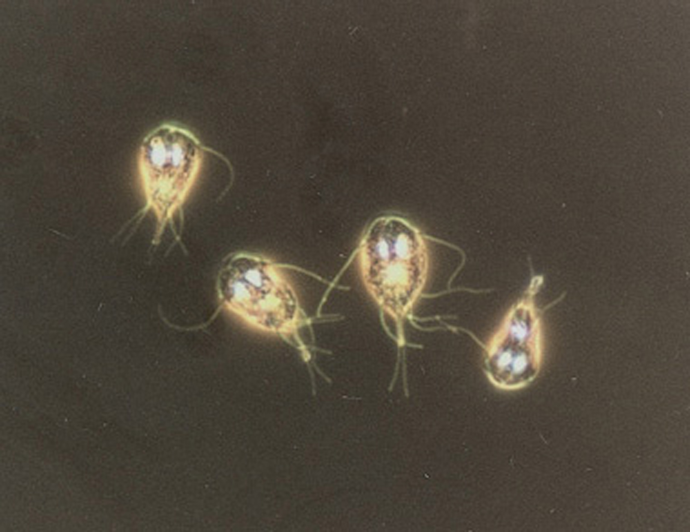
| 2 september 2011 kl. 13.35 | GiardiaTrofozAM.jpg (fil) |  |
334 kbyte | Foto: Anders Magnusson, Smittskyddsinstitutet | 1 |
| 19 april 2012 kl. 16.09 | G 100x.jpg (fil) |  |
277 kbyte | Toxoplasma /monocyter, Giemsa. Foto Silvia Botero Kleiven | 1 |
| 20 september 2011 kl. 14.08 | H.diminutaÄggML-a.jpg (fil) |  |
283 kbyte | Foto:marianne Lebbad, Smittskyddsinstitutet | 1 |
| 28 mars 2012 kl. 09.59 | HakmaskÄggAM.jpg (fil) |  |
451 kbyte | Hakmask-ägg. Foto: Anders Magnusson, SMI | 1 |
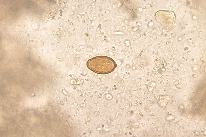
| 8 september 2011 kl. 16.12 | Heterophyes egg.jpg (fil) |  |
64 kbyte | Foto: CDC via Wikimedia Commons | 1 |
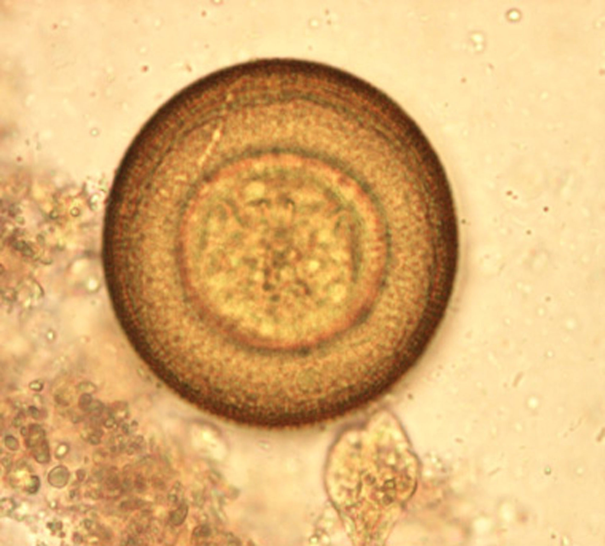
| 15 september 2011 kl. 15.14 | H nana adultF. cdc.jpg (fil) |  |
15 kbyte | CDC via Wikimedia Commons | 1 |
| 15 september 2011 kl. 15.16 | H nana eggB. G.dept.publ.health.jpg (fil) |  |
18 kbyte | Georgia Dept. Publ. Health via Wikimedia Commons | 1 |
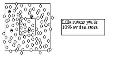
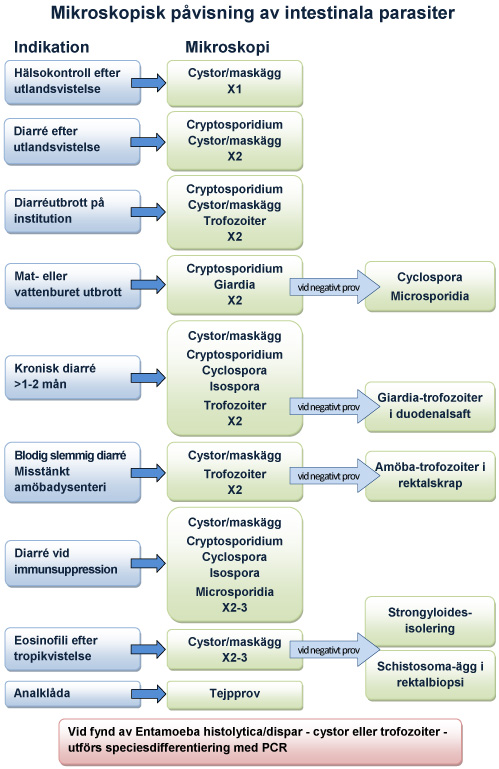
| 11 april 2012 kl. 12.28 | IndikationMikroskopi.jpg (fil) |  |
176 kbyte | Påvisning av intestinala parasiter | 1 |
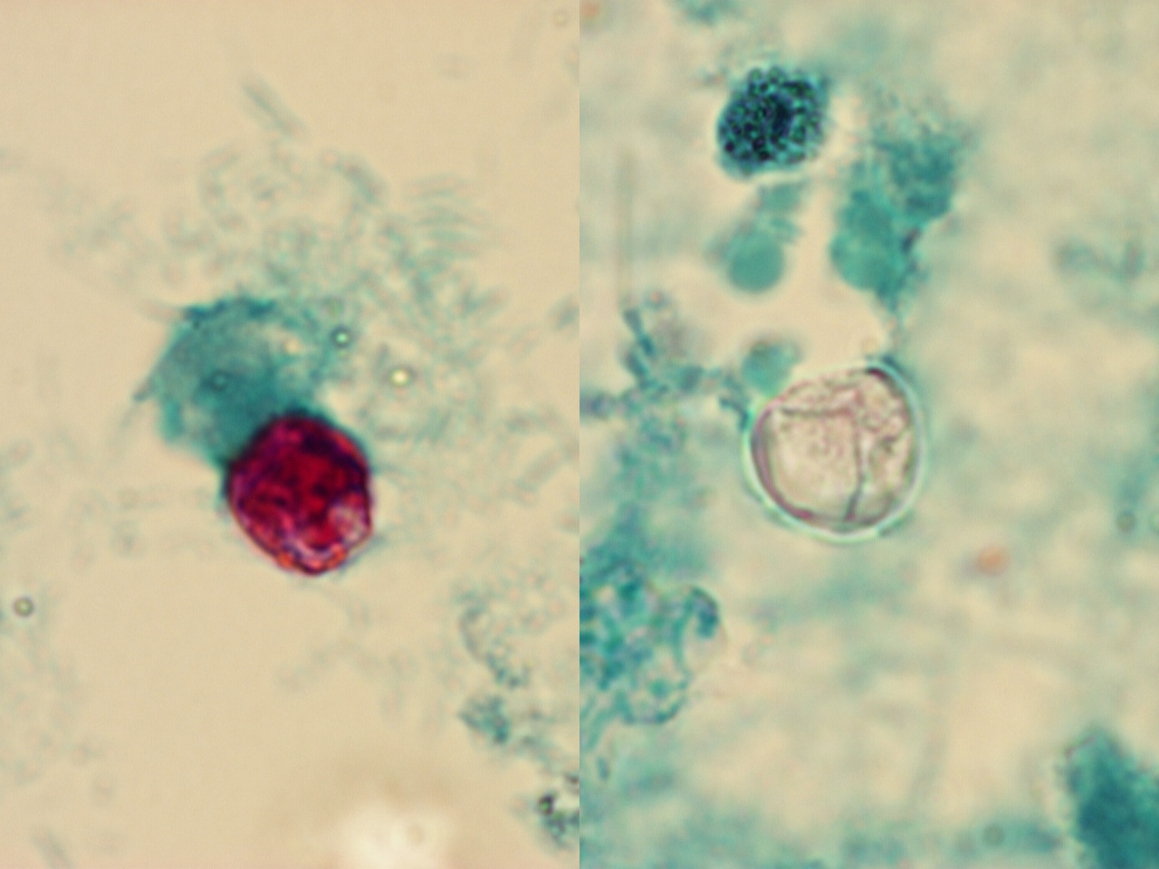
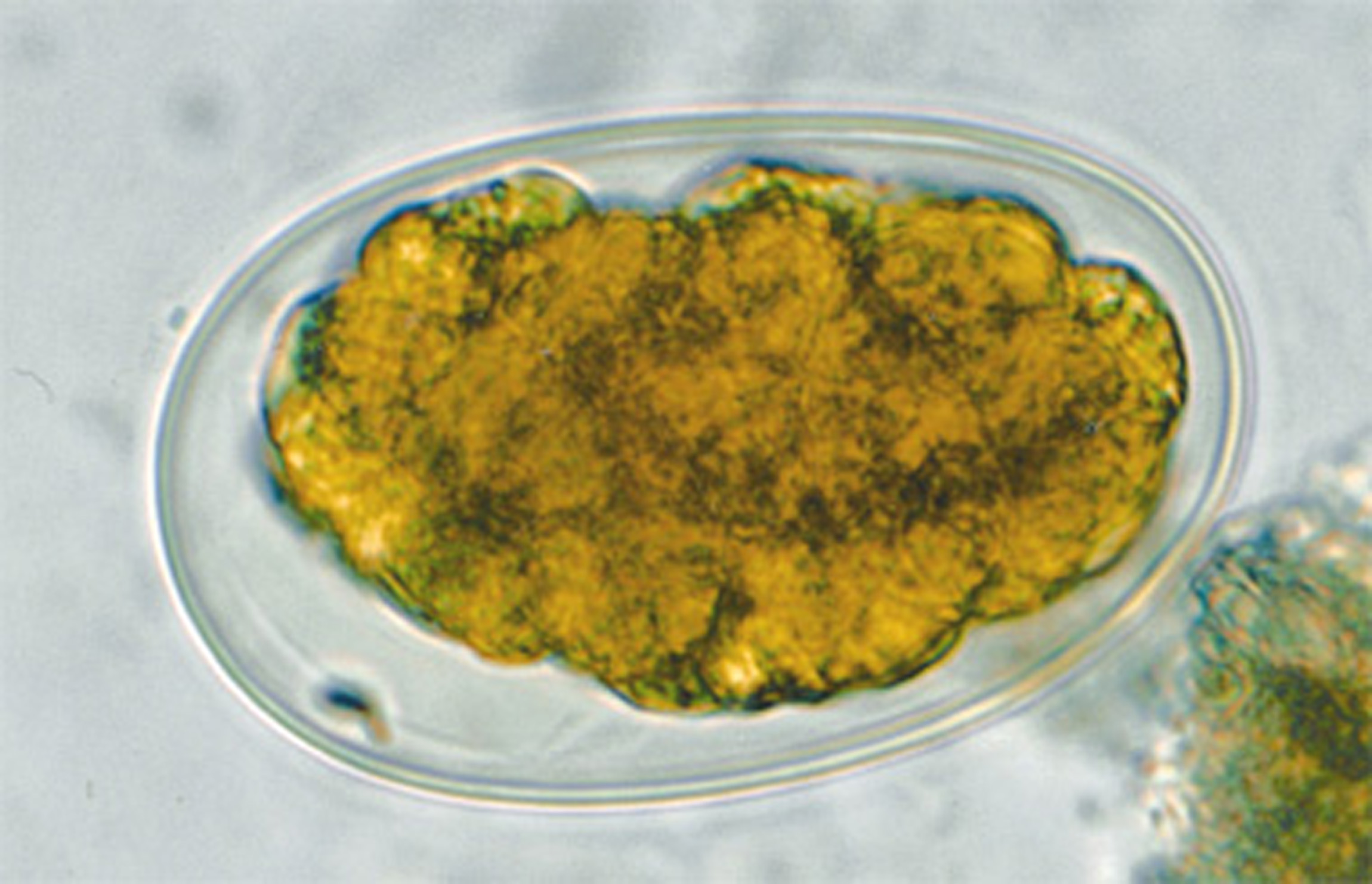
| 6 mars 2012 kl. 15.32 | Isospora bild 2.jpg (fil) |  |
675 kbyte | OOcysta från Isospora (Cycloisospora) hominis. Foto: Marianne Lebbad SMI | 1 |
| 21 augusti 2012 kl. 12.24 | Isospora ML.jpg (fil) |  |
6 kbyte | Isospora, Z-N färgning, foto Marianne Lebbad, Smittskyddsinstitutet | 1 |
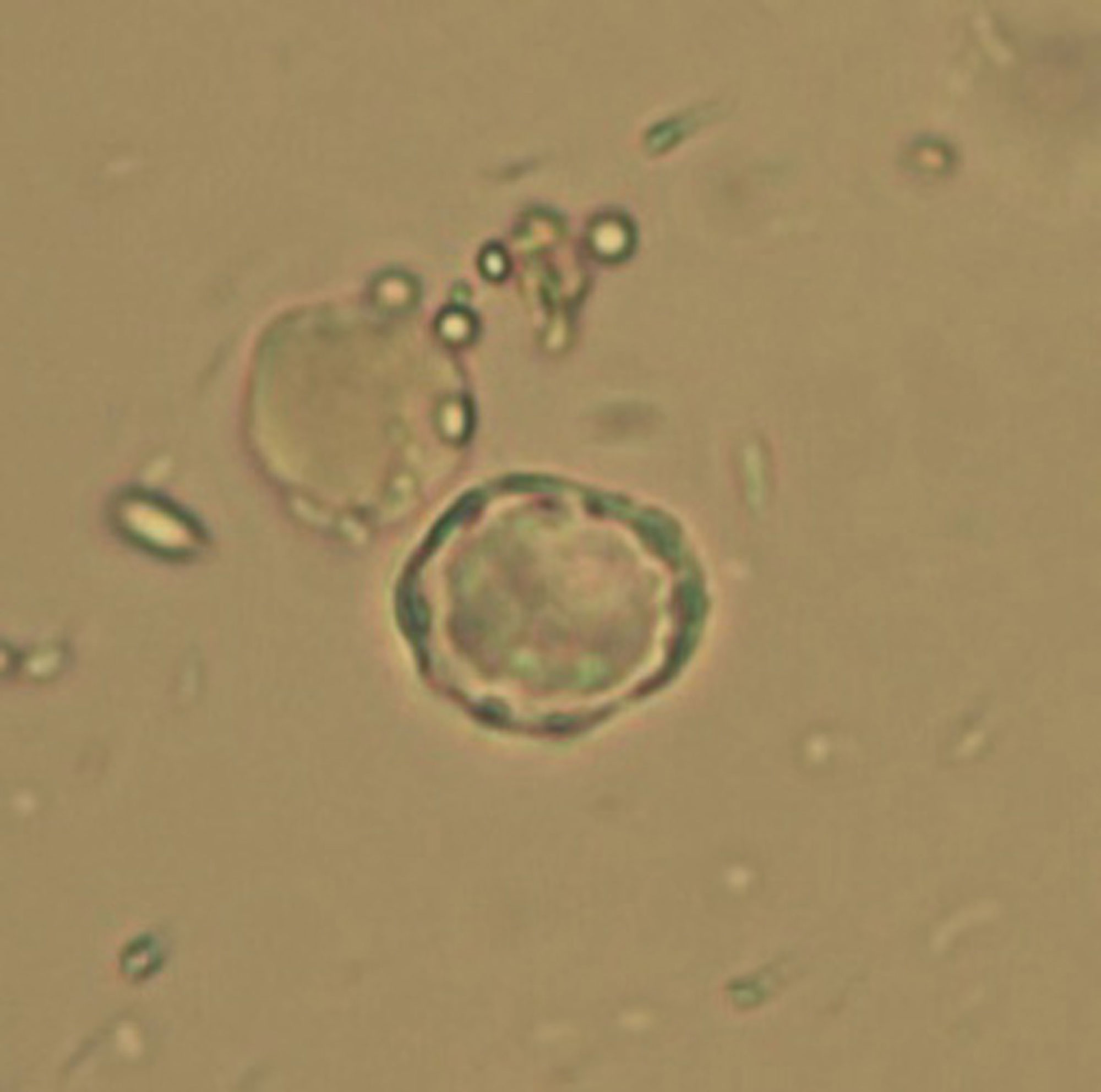
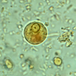
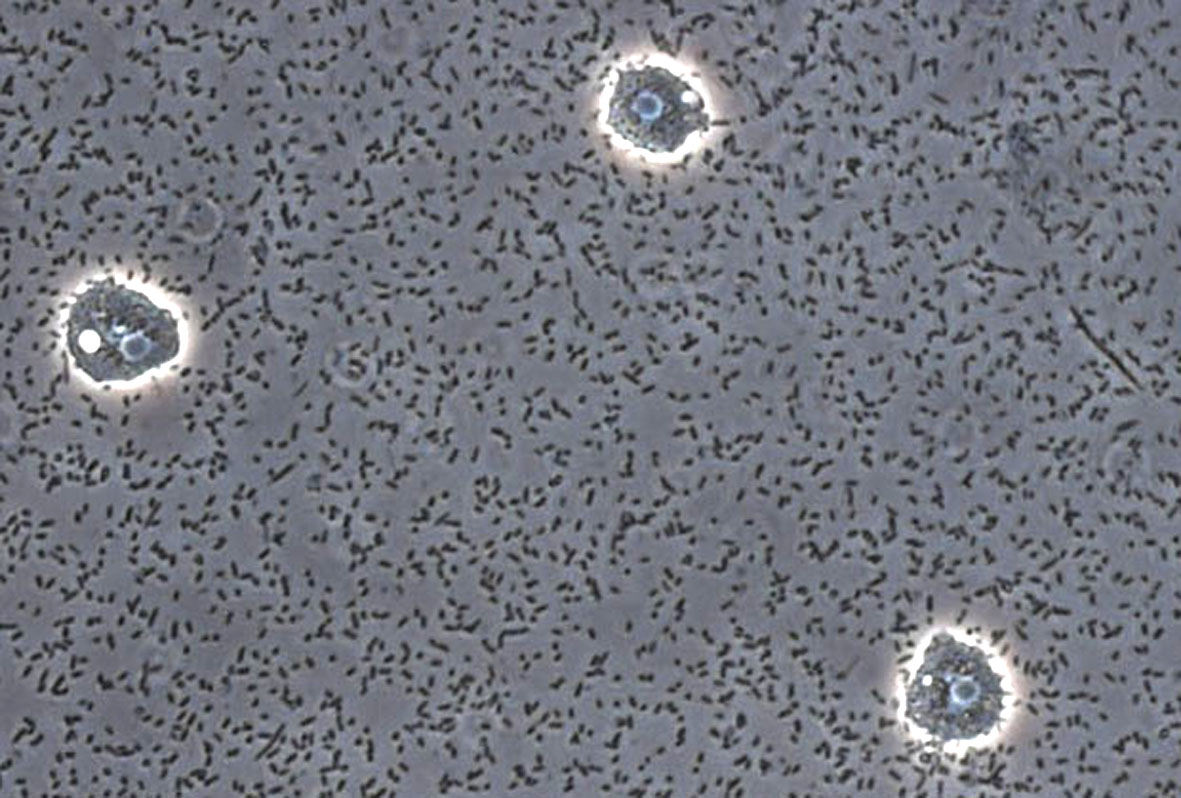
| 19 augusti 2011 kl. 11.59 | Jadwiga Acanthamoeba 40 obj. .0006.jpg (fil) |  |
207 kbyte | Acanthamoeba. Foto Jadwiga Krusnell | 1 |
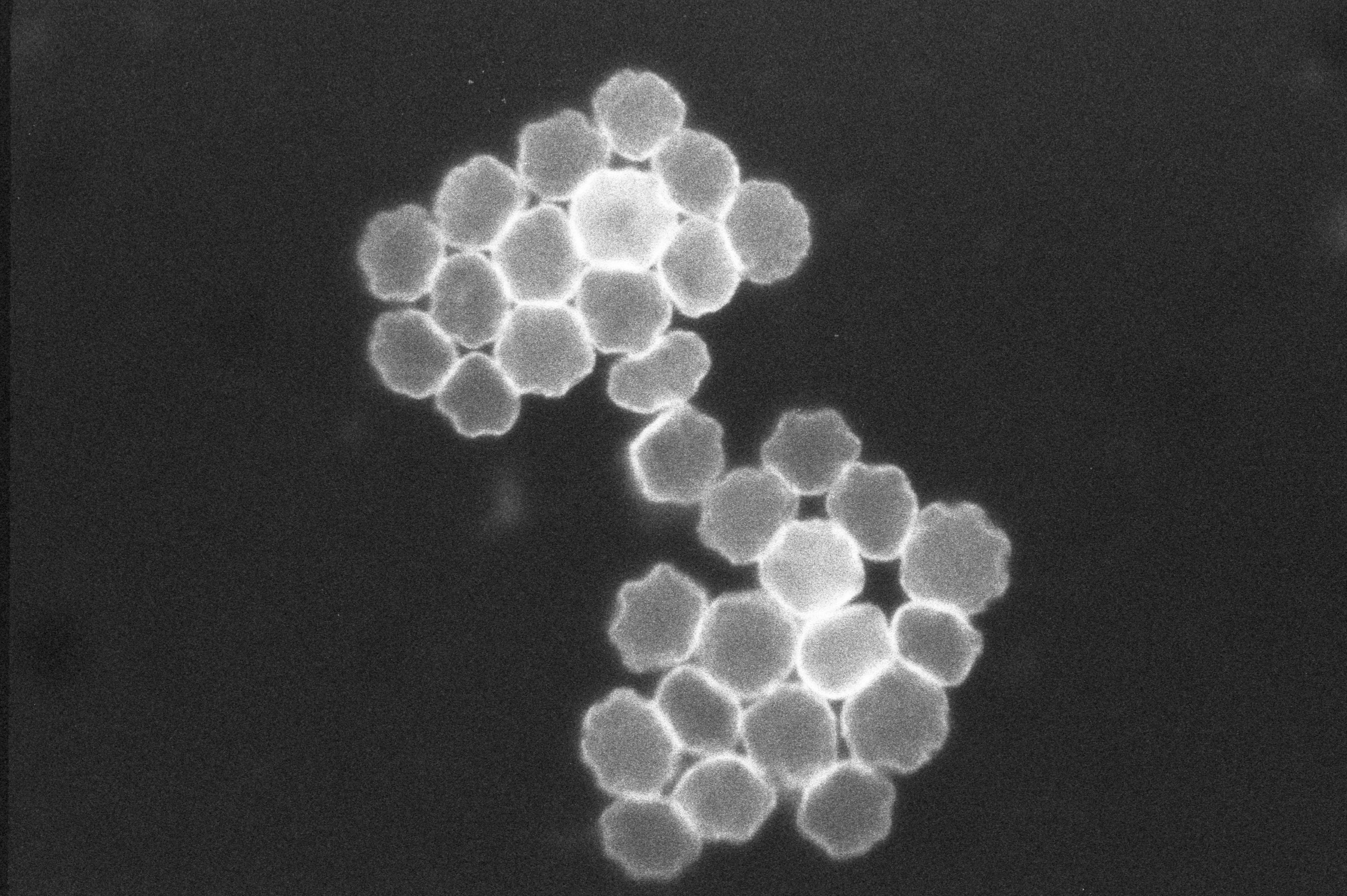
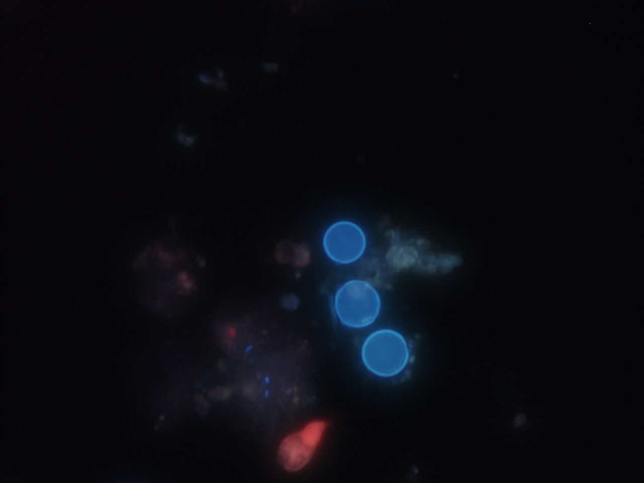
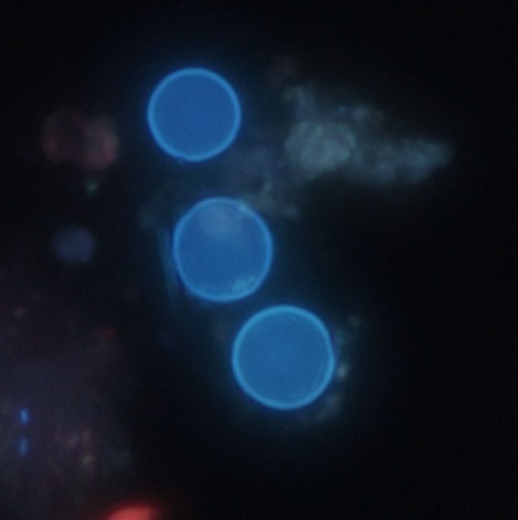
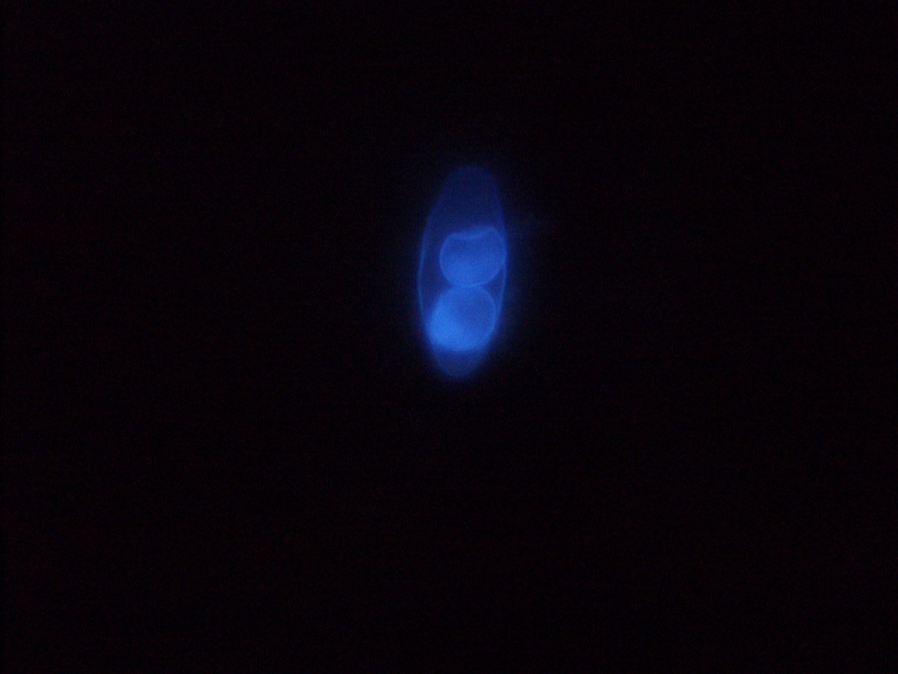
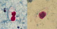
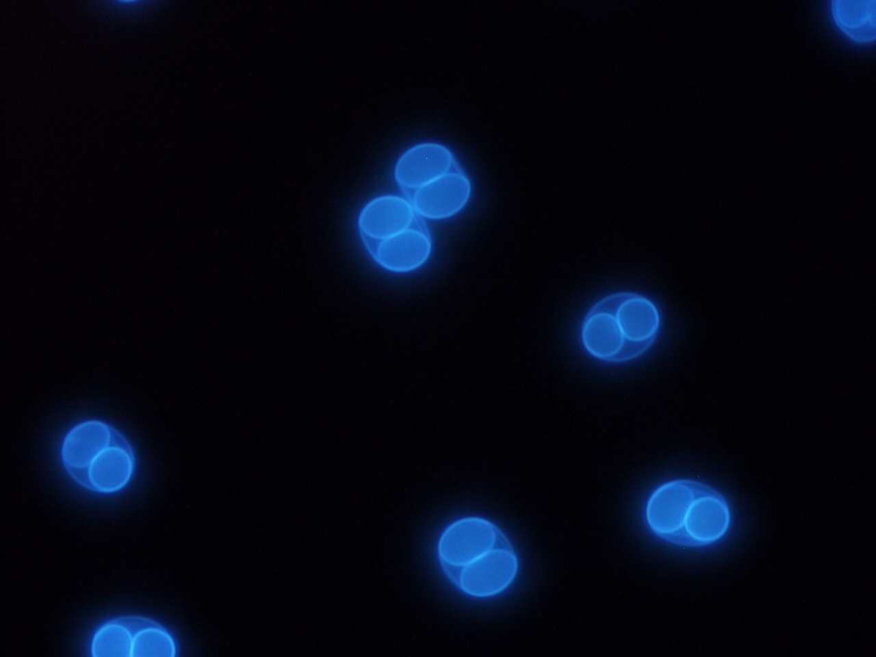
| 26 augusti 2011 kl. 09.16 | Jadwiga Toxo oocysts.jpg (fil) |  |
590 kbyte | Autofluorescerande oocystor av T. gondi | 1 |
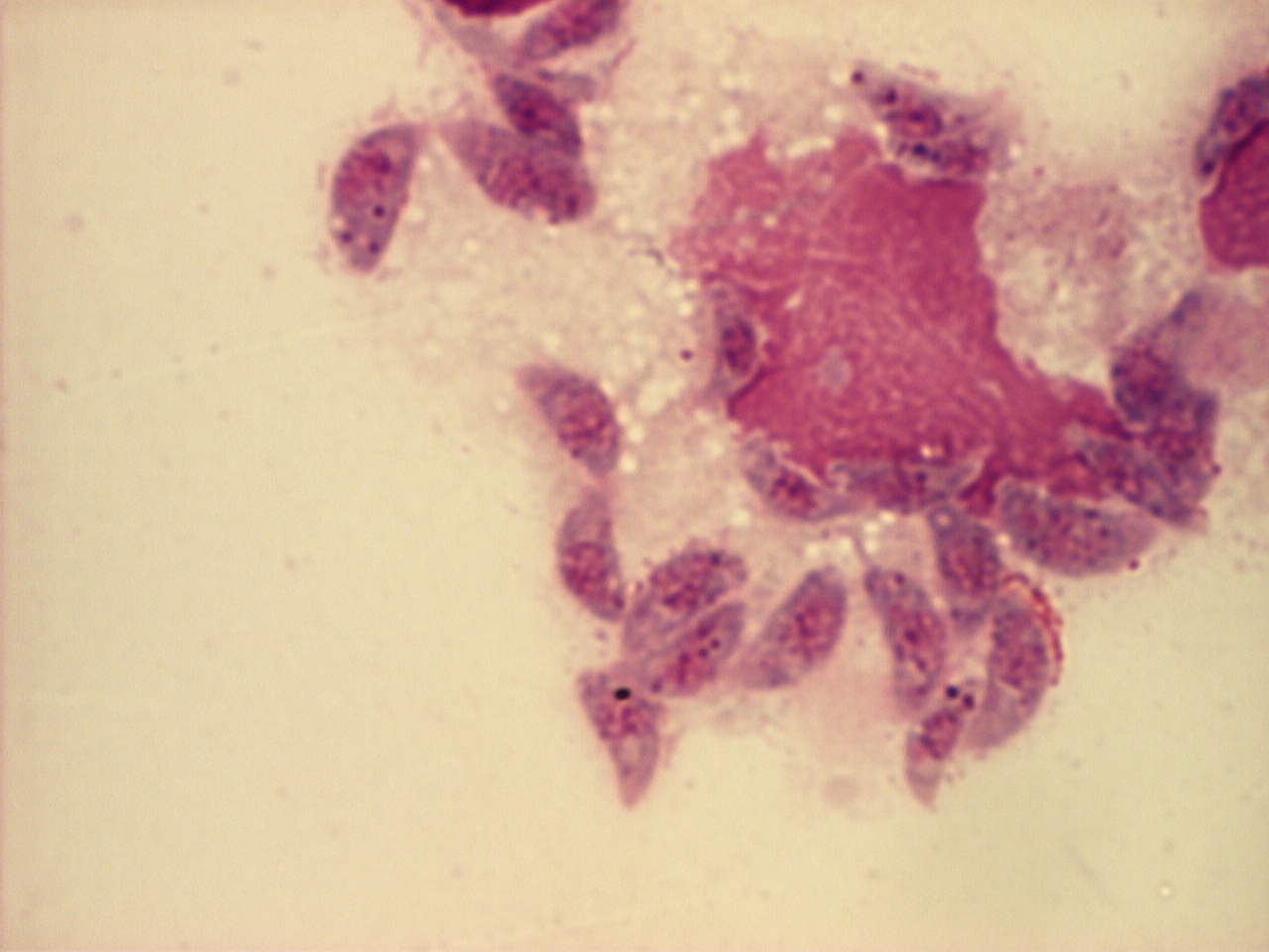
| 19 april 2012 kl. 16.07 | J 100x.jpg (fil) |  |
284 kbyte | T. gondii/monocyter, Giemsa. Foto Silvia Botero Kleiven | 1 |
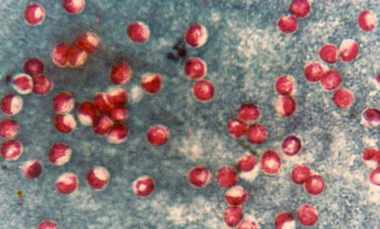
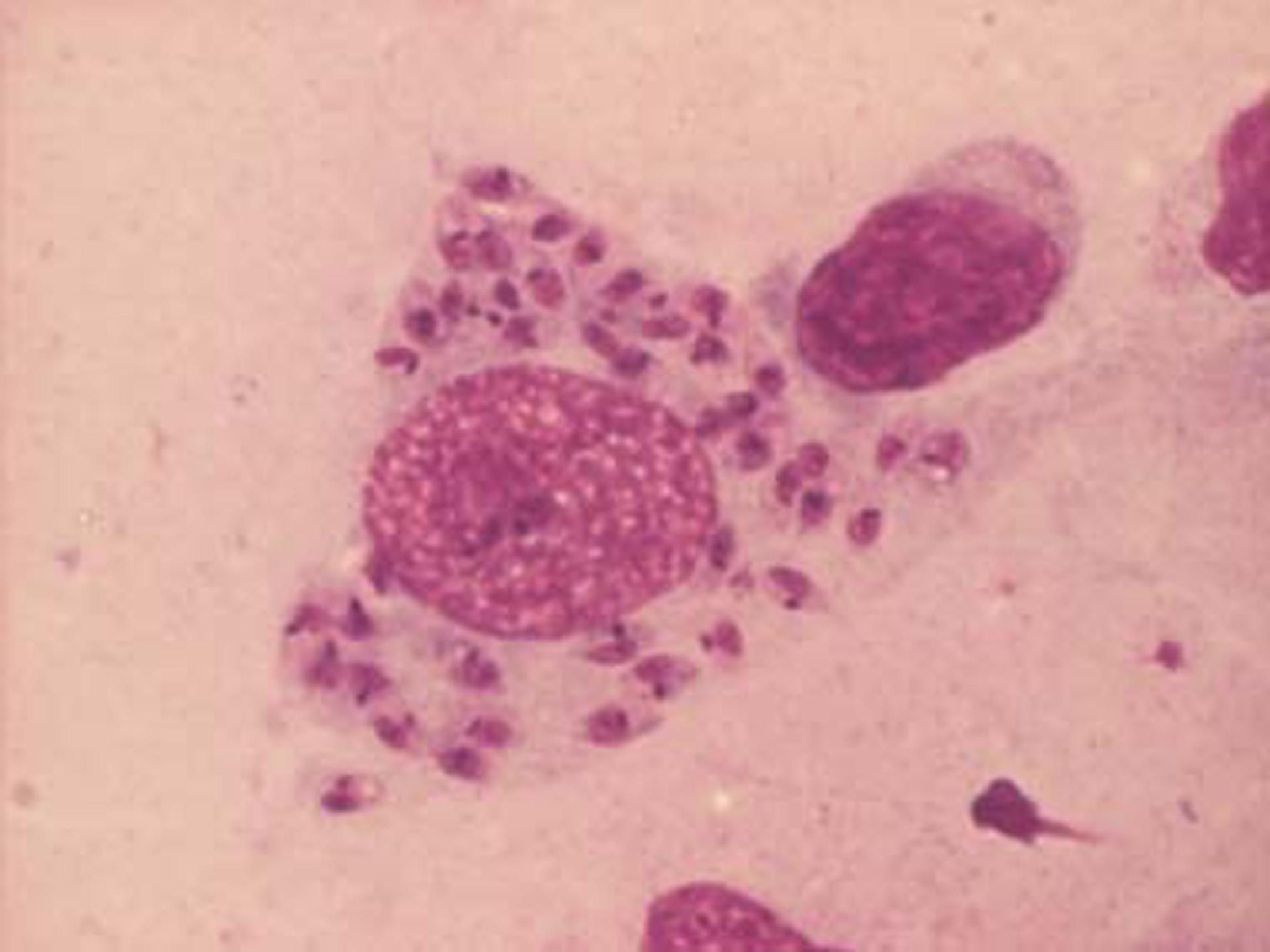
| 26 augusti 2011 kl. 15.45 | LeishmAmastigML.jpg (fil) |  |
275 kbyte | Foto: Marianne Lebbad, Smittskyddsinstitut | 1 |
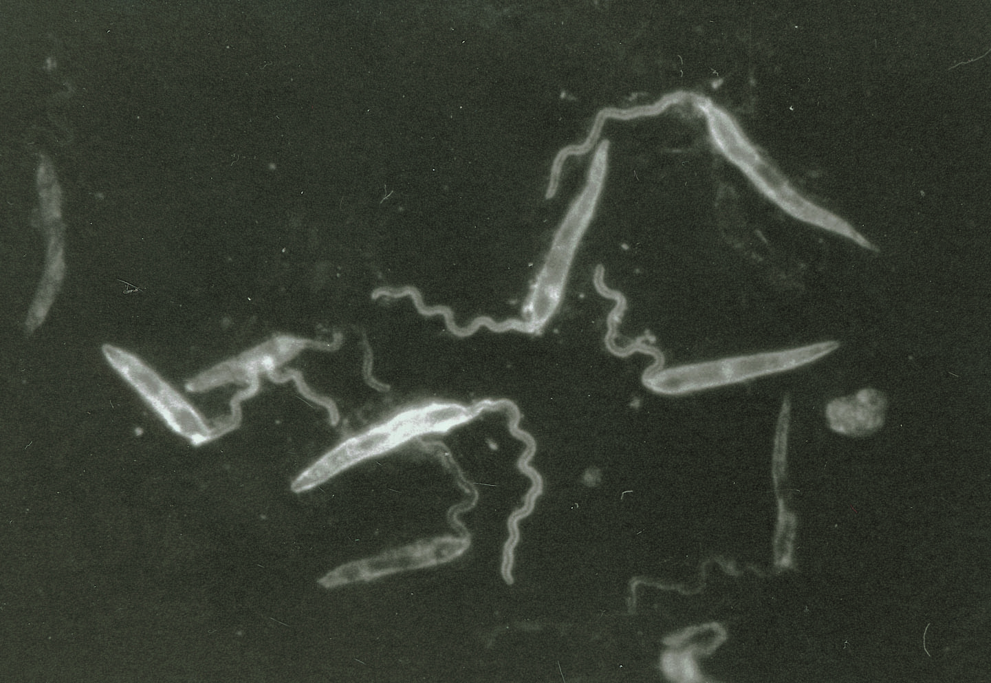
| 19 augusti 2011 kl. 11.55 | Leishmania1.jpg (fil) |  |
319 kbyte | Leishmania promastigotes i RPMI. Foto Jadwiga Krusnell | 1 |
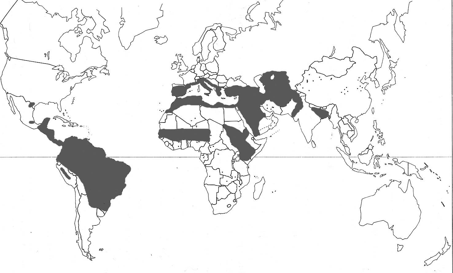
| 19 augusti 2011 kl. 11.36 | Leishmaniakarta 3.jpg (fil) |  |
120 kbyte | 1 | |
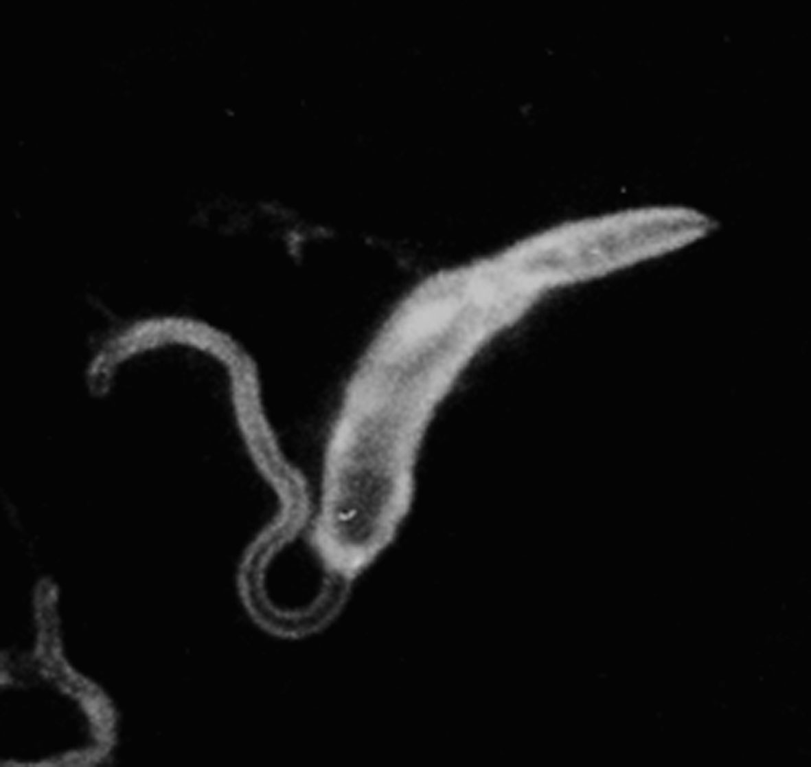
| 26 augusti 2011 kl. 12.22 | Leishmaniapromastigote.jpg (fil) |  |
61 kbyte | Foto: Jadwiga Winiecka-Krusnell, Smittskyddsinstitutet | 1 |